


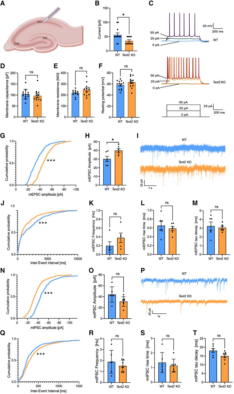
Tent2 KO neurons exhibit increased excitability in CA1 in hippocampal slices (A–F) and enhanced excitatory synaptic transmission in cultured hippocampal neurons (G–M). (A) Schematic of patch-clamp recordings in CA1 region of hippocampal horizontal slices. (B) Rheobase, the lowest value of current to trigger an action potential was significantly lower for Tent2 KO (Mann–Whitney test; [*] P = 0.0257). (C) Representative voltage responses to current stimuli for WT (top panel) and Tent2 KO neurons (middle panel). (D–F) Statistics for selected passive membrane properties (see Materials and Methods for description). Membrane capacitance: membrane resistance or resting membrane potential were not altered in Tent2 KO neurons. Data were obtained from 12 to 13 neurons per group. The data are expressed as mean ± SEM. (G–M) AMPA/kainate receptor glutamatergic transmission was recorded in 21–22 DIV hippocampal cultures with whole-cell patch-clamp method. (G) The cumulative probability of individual mEPSCs amplitudes was shifted toward higher values (Kolmogorov–Smirnov test; P < 0.00001). (H) Averaged amplitude of mEPSCs was significantly larger for Tent2 KO neurons (t-test; P < 0.05). (I) Representative current traces from WT and Tent2 KO neurons. (J) The cumulative distribution function for inter-event time intervals between subsequent mEPSCs is left-shifted compared to WT neurons (Kolmogorov–Smirnov test P < 0.0001). (K) The averaged frequency of mEPSCs is not significantly changed between genotypes. (L and M) Tent2 KO neurons do not exhibit altered mEPSCs rise time (L) or tau decay (M). Data were obtained from six neurons per group. The data are expressed as mean ± SEM. (N–T) Reduction of inhibitory synaptic transmission in Tent2 KO cultured hippocampal neurons. (N) The cumulative probability of amplitudes for mIPSCs is left-shifted (Kolmogorov–Smirnov test; P < 0.0001). (O) Averaged amplitudes of mIPSCs were not significantly different in Tent2 KO neurons (t-test; P = 0.07). (P) Representative current traces of mIPSCs recordings from WT and Tent2 KO cultured neurons (21–22 DIV). (Q) The cumulative probability plot of the inter-event interval for Tent2 KO is right-shifted relative to the WT neurons (Kolmogorov–Smirnov test P < 0.0001). (R) The averaged frequency of mIPSCs was not significantly changed. (S and T) mIPSCs rise time (S) and tau decay (T) were not altered in Tent2 KO neurons. Data were obtained from six to seven neurons per group. The data are expressed as mean ± SEM. Partially created with Biorender.




