


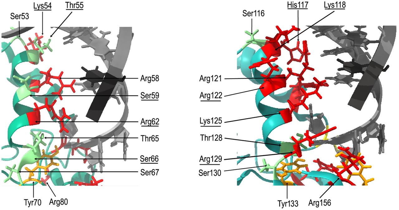
The modeling of RNA-binding surfaces of E. coli ProQ and F-like plasmid FinO. The figure shows the α-helix 3 and surrounding region from E. coli ProQ (left) and the corresponding α-helix 4 from F-like plasmid FinO (right) with those amino acid residues marked, which side chains are directed toward the modeled location of RNA helix. The modeling of interactions was done using Chimera X (Pettersen et al. 2021) by aligning the NMR structure of the FinO domain of E. coli ProQ (Gonzalez et al. 2017) and the X-ray structure of the F-like plasmid FinO protein (Ghetu et al. 2000) with the X-ray structure of the FinO domain of L. pneumophila RocC in complex with the terminator hairpin of RocR RNA (Kim et al. 2022). The side chains of amino acid residues located in the corresponding positions of both proteins are marked in color, with arginine, lysine, and histidine residues marked in red, serine and threonine in green, and tyrosine in orange. The descriptions of corresponding amino acids are located in corresponding places on the figure. Those amino acids which are different, but located in corresponding positions, are marked by underlining. The structure of the L. pneumophila RocR hairpin is shown in gray.




