


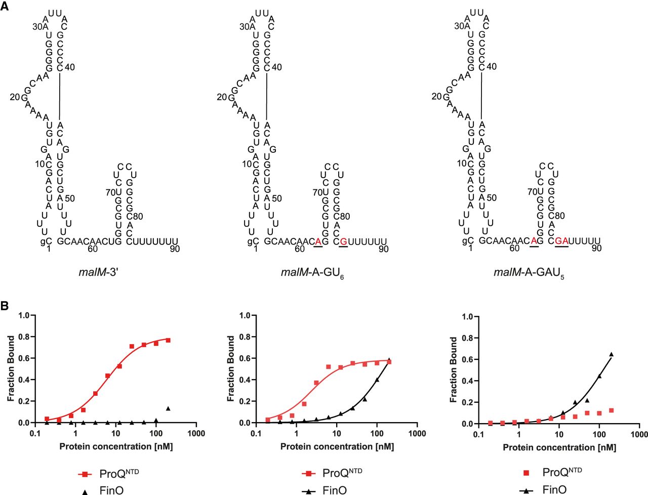
Comparison of malM-3′, malM-3′-A-GU6, and malM-3′-A-GAU5 binding to ProQNTD, and to FinO. (A) Secondary structures of malM-3′, malM-3′-A-GU6, and malM-3′-A-GAU5, which were predicted using RNAstructure software (Reuter and Mathews 2010). The nucleotides from FinP which were substituted into malM-3′ are shown in red underlined font. The lowercase g denotes guanosine residue added on 5′ ends of RNA molecules to enable T7 RNA polymerase transcription. (B) The respective binding data for ProQNTD and FinO are shown on the graphs below each RNA. The fitting of the quadratic equation into malM-3′-A-GU6 data provided a Kd value of 1.8 nM for binding to ProQNTD, and 151 nM for binding to FinO. The fitting of the quadratic equation into malM-3′-A-GAU5 data provided a Kd value of 122 nM for binding to FinO, while the Kd value for binding to ProQNTD was estimated as higher than 200 nM. The data shown for malM-3′ are the same as in Figure 1. Gels corresponding to the data in the plots are shown in Supplemental Figure S7. Average Kd values are shown in Table 1.




