


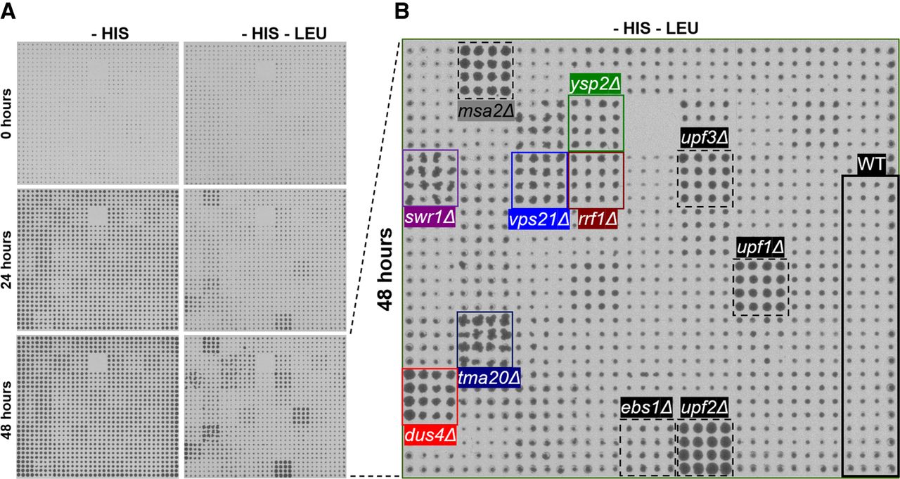
FIGURE 3.
Finalized qualitative screen using pTDH3_HIS3_PGA1. (A) Image at the start of the experiment and at 24 and 48 h intervals are shown. (B) Image at the end of the experiment under the selective condition at 48 h is shown (magnification of bottom right-hand field in A. Mutants affected in previously known NMD factors are highlighted (black), as well as candidates that show strong phenotype and were used for further experiments: ysp2Δ (green), swr1Δ (purple), vps21Δ (blue), rrf1Δ (brown), tma20Δ (dark blue), dus4Δ (red). Colonies marked in stippled black represent a misplaced strain (msa2Δ) and were ignored.




