


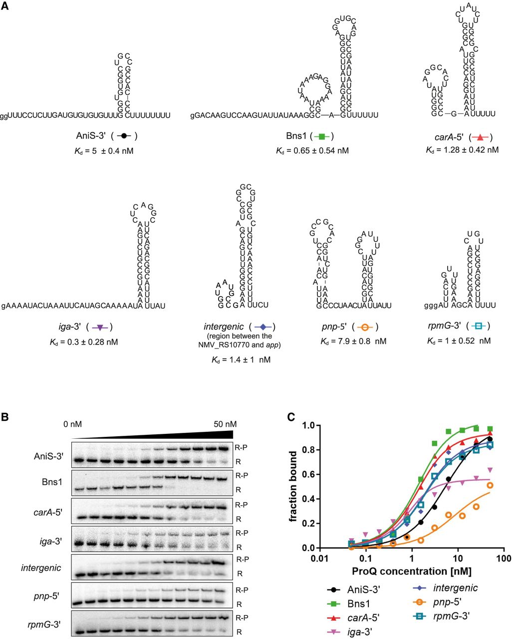
RNA molecules bound by ProQ protein in N. meningitidis, which were used in this study. (A) The secondary structures of RNAs AniS-3′, Bns1, carA-5′, iga-3′, intergenic (intergenic region between NMV_RS10770::app), pnp-5′, and rpmG-3′. The lower-case g denotes guanosine residue added on 5′ end to enable T7 RNA polymerase transcription. (B) The binding of 32P-labeled RNAs AniS-3′, Bns1, carA-5′, iga-3′, intergenic, pnp-5′, and rpmG-3′ to ProQ was monitored using a gel-shift assay. Free 32P-RNA is marked as R, RNA-ProQ complexes as R-P. (C) The fitting of the ProQ binding data from B using the quadratic equation provided Kd values of 4.9 nM for AniS-3′, 0.9 nM for Bns1, 0.9 nM for carA-5′, 0.3 nM for iga-3′, 1.4 nM for intergenic, 7.9 nM for pnp-5′, and 1.2 nM for rpmG-3′. The RNA secondary structure predictions were performed in the ViennaRNA program (Lorenz et al. 2011). Raw gel data for all RNAs are presented in Supplemental Figure S3. The average equilibrium dissociation constant (Kd) values and the maximum RNA fraction bound calculated from at least three independent experiments are shown in Table 1.




