


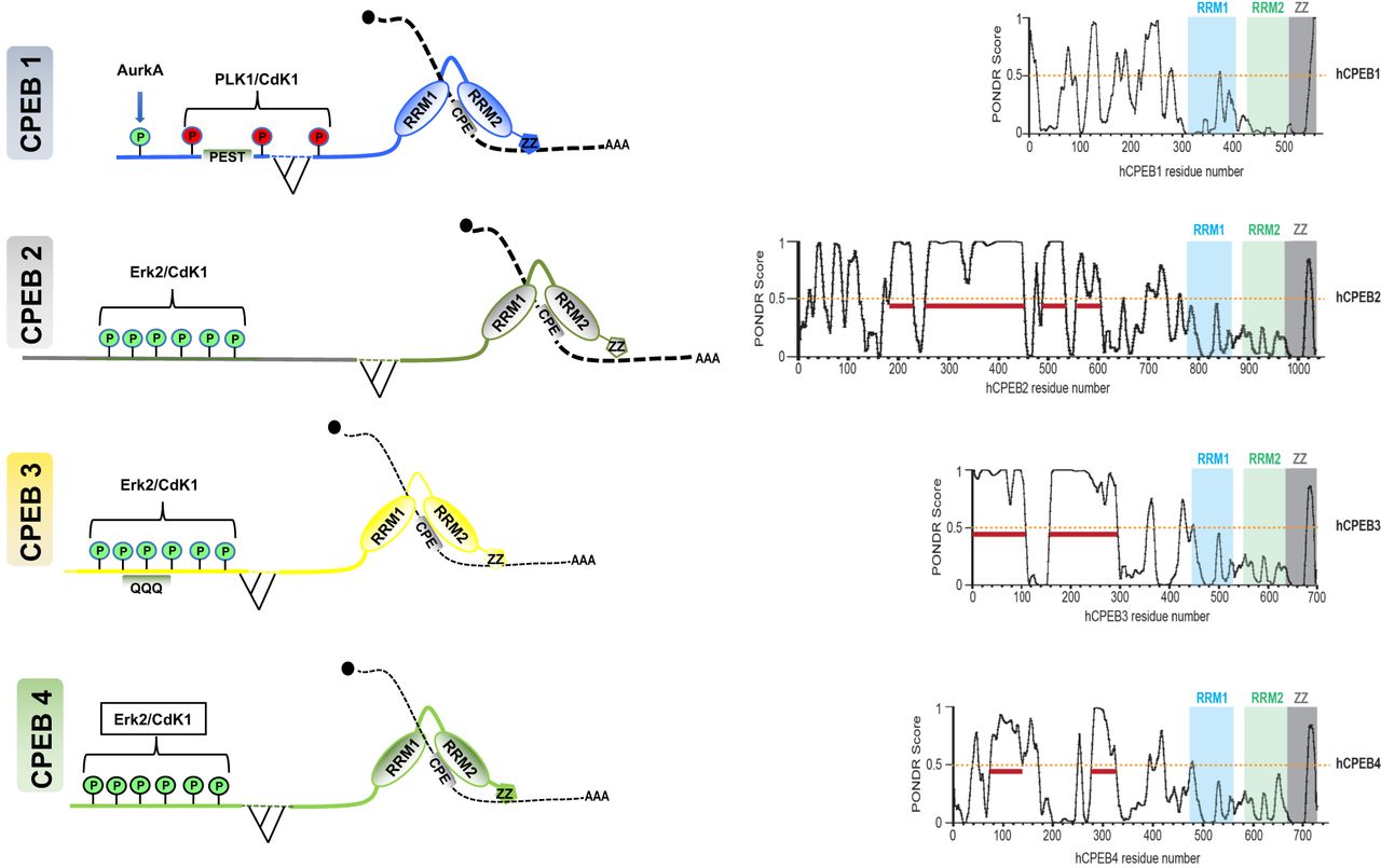
CPEB domains and regulatory phosphorylations. (Left) All four CPEBs share a similar structure, with a highly ordered C-terminal domain comprising two RRMs in tandem and a ZZ-box responsible for binding to their targets, and a highly variable NTD, which contains regulatory phosphorylation sites (activating sites in green, repressing sites in red; kinases indicated). CPEBs can also undergo alternative splicing, generating isoforms with potentially different behaviors and functions. (Right) CPEBs 2–4 are predicted to be intrinsically disordered and contain low-complexity regions (LCRs) within their variable NTDs. POND-score profiles of CPEBs 1–4 show stretches of predicted disordered regions.




