


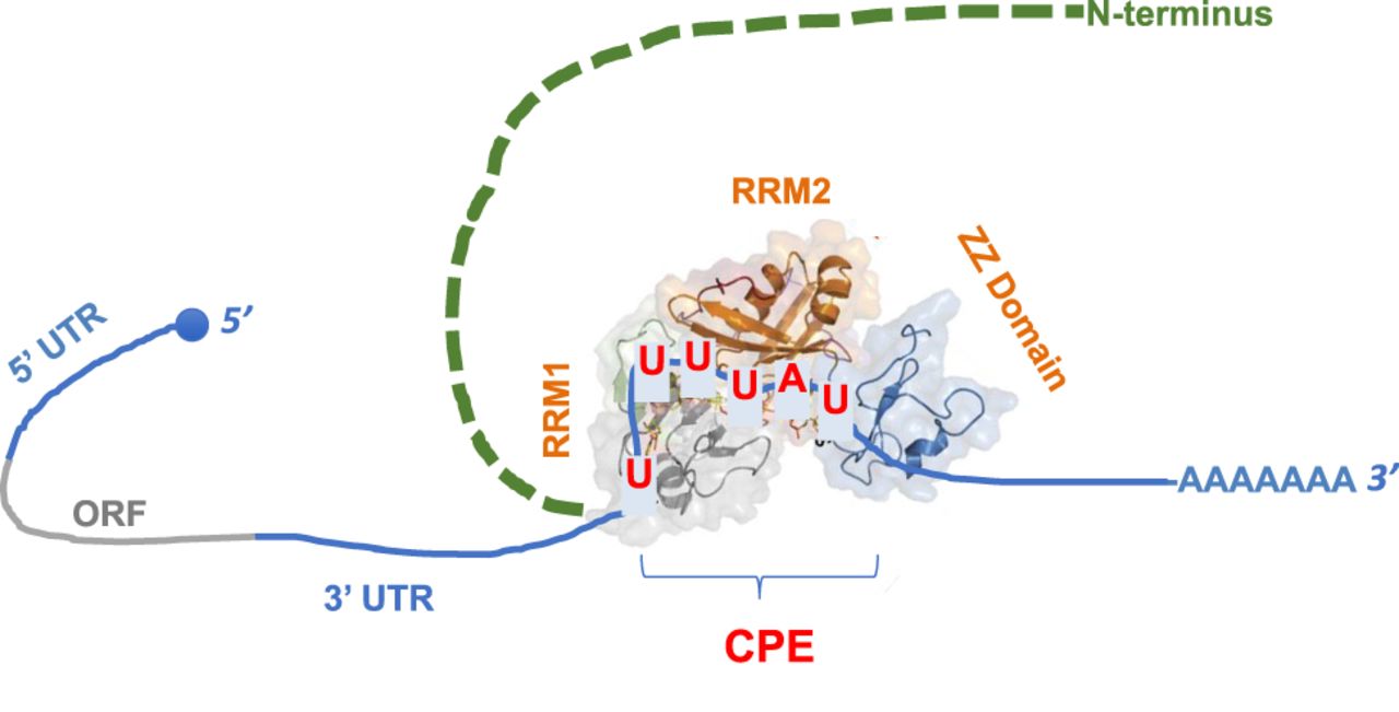
FIGURE 1
. Schematic representation of a CPEB bound to the CPE in the 3′ UTR of the target mRNA. The structure of the two RRMs and the ZZ-box is adapted with permission from Afroz et al. (2014). The intrinsically disordered N-terminal half of CPEBs is indicated, along with the 5′ and 3′ UTRs of the mRNA, the open reading frame (ORF), and the poly(A) tail.




