


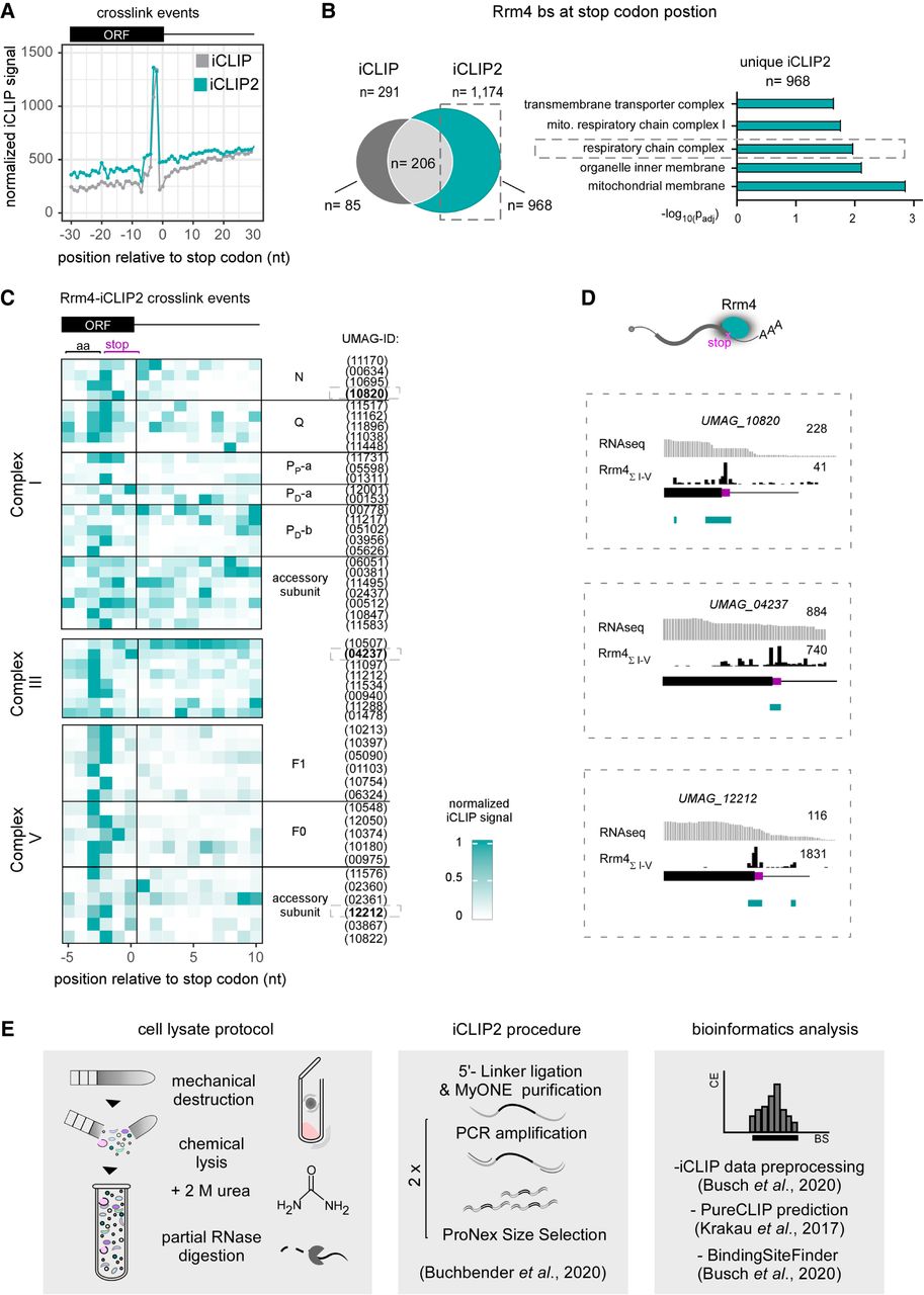
Rrm4 binds at stop codons of nuclear-encoded mRNAs of the respiratory chain complex I, III, and V. (A) Metaprofile of Rrm4 iCLIP (gray) and iCLIP2 (petrol) normalized signals relative to stop codon. (B) Venn diagram showing the number of Rrm4-binding sites at the stop codon position as detected by iCLIP and iCLIP2 (dark gray: uniquely found in iCLIP n = 85; light gray: shared targets n = 206; petrol: uniquely found in iCLIP2 n = 968). Selected bar chart from GO-term analysis of stop codon binding targets uniquely determined within the iCLIP2 data set. (C) Heatmap of Rrm4 iCLIP2 cross-link events in a window (−5 to 10 nt) around the stop codon position (0 is defined as the last nucleotide of the stop codon). The amino acid codon preceding the stop codon is indicated (aa). The core complex subunits N, Q, and P, as well as the accessory subunits of respiratory complex I, are indicated. Complex III and the subunits of complex V (F1 and F0) and their accessory subunits are also indicated. The respective identifiers (UMAG_ID) of the individual proteins are listed alongside. (D) Genome browser views of nuo2 (UMAG_10820, Comp I), qcr7 (UMAG_04237, Comp III), and atp10 (UMAG_12212, Comp V) mRNAs. The predicted binding sites are depicted in petrol, and the stop codon is highlighted in magenta. (E) Schematic overview of the optimized microbial iCLIP2 procedure. Lysis protocol (left) was improved by the adaption of dry-cryogenic milling, buffer optimization by adding the chaotropic detergents urea (2 M), and internal RNase digestion. The adaptation of the advanced iCLIP2 procedure (center) and defined bioinformatics analysis (right), including data preprocessing, PureCLIP, and BindingSiteFinder is indicated (described in Materials and Methods).




