


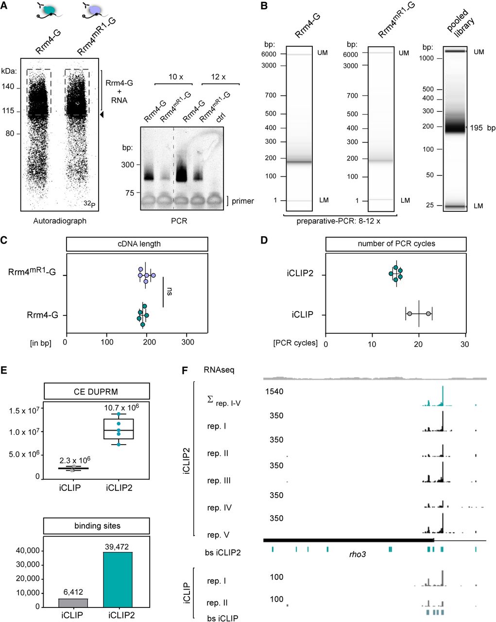
Adaption of improved iCLIP2 protocol. (A) Autoradiograph and optimization-PCR of Rrm4/Rrm4mR1-G. (B) Capillary gel electrophoresis image of the final PCR of Rrm4/Rrm4mR1-G libraries before and after pooling. The respective number of PCR cycles depends on the RBP (Rrm4 vs. Rrm4mR1-G) and the quantity of the respective cDNA library. The pooled library includes additional samples beyond Rrm4-G. (C) Reproducible cDNA length based on the mean of n = 5 biological replicates of Rrm4-G/Rrm4mR1-G. (D) Number of PCR cycles for Rrm4-G needed for library preparation by iCLIP (18–22×; n = 2) or iCLIP2 (1. PCR: 6×; 2. PCR: 8–10×; in total = 14–16×; n = 5) procedure. (E) Quantified numbers of cross-link events after duplicate removal (CE after DUPRM) of Rrm4-G iCLIP (n = 2) and Rrm4-G iCLIP2 (n = 5). Number of binding sites determined for Rrm4-G in iCLIP (n = 6412) versus iCLIP2 (n = 39,472). For both data sets, a nucleotide window of 9 nt has been chosen. (F) Genome browser view of rho3 mRNA. The numbers represent the upper limit of stack heights for iCLIP(2) cross-link events (CE) and RNA-seq reads. The RNA-seq coverage of Wt filaments is depicted in gray. The five biological Rrm4 replicates of iCLIP2 are depicted in black (mentioned as rep. I–V). The respective merged cross-link events of all five biological replicates and the determined binding sites (bs) are depicted in petrol. The rho3 ORF is shown as a filled rectangle. The two biological Rrm4 iCLIP replicates (gray, rep. I/II) and the determined bs (light petrol) are depicted underneath.




