


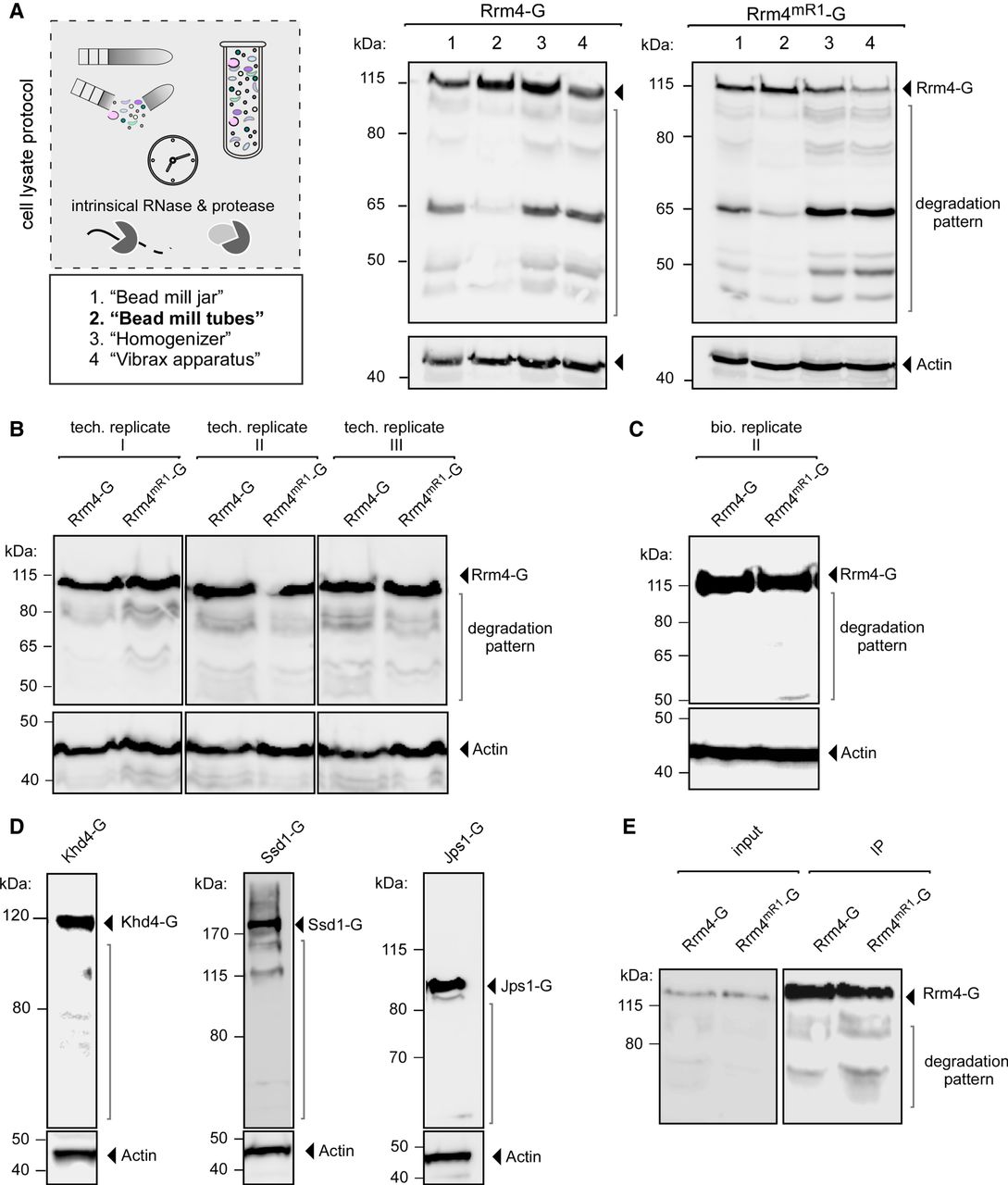
Improved mechanical cell distribution. (A) Four techniques to mechanically lyse cells were analyzed (1 = Bead mill jar, 2 = Bead mill tubes, 3 = Homogenizer, 4 = Vibrax apparatus). Start material from the same batches (Rrm4/Rrm4mR1-G) was used. The respective cell lysates were subjected to western blot analysis (Rrm4/Rrm4mR1-G = 112 kDa, indicated by arrow). The area of protein degradation is marked in dark gray (Actin, 42 kDa, served as control). (B) Western blot analysis of three technical replicates of technique 2 (Bead mill tubes) are shown. Technical replicates of Rrm4/Rrm4mR1-G cell lysates were prepared from the same batches of frozen materials. The full-length protein is indicated by an arrow (112 kDa), and the respective degradation patterns are marked (Actin, 42 kDa, served as control). (C) Western blot analysis as in B. The respective biological replicates were performed as described previously. (D) Western blot analysis of the RBPs Khd4-G (178 kDa), Ssd1-G (180 kDa), and the protein Jps1-G (90 kDa; actin, 42 kDa, served as control). (E) Western blot analysis of immunoprecipitation of Rrm4/Rrm4mR1-G (IP, 112 kDa, indicated by arrow).




