


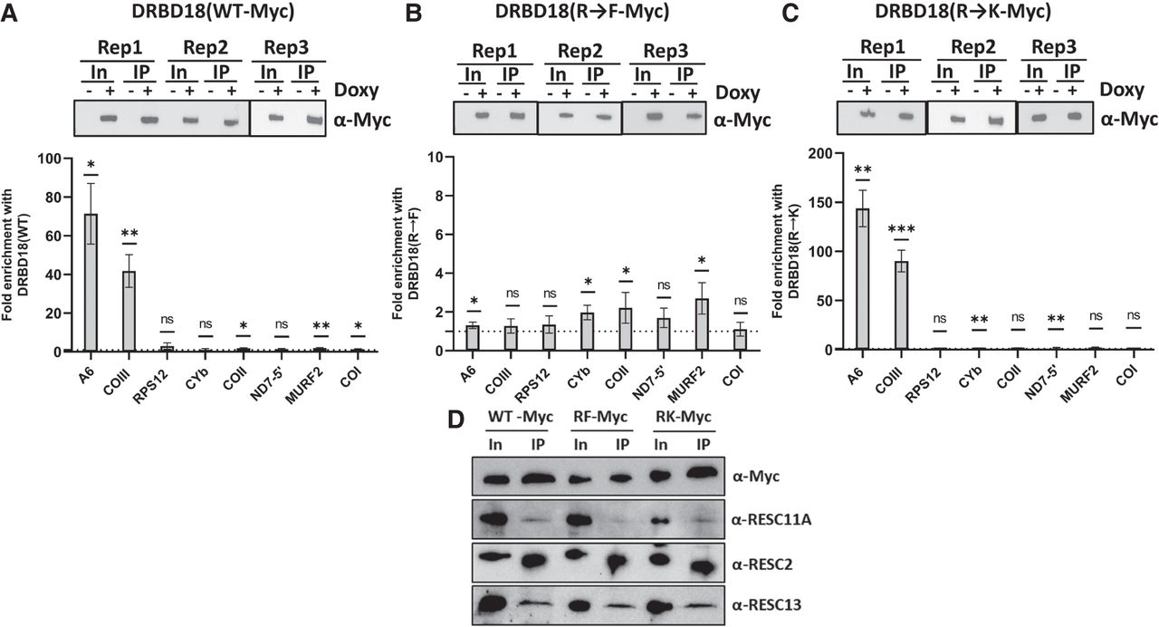
Association of wild type and methyl mutant DRBD18 with mitochondrial transcripts and RESC proteins. (A, top) Western blot showing comparable DRBD18(WT) IP in three biological replicates. In, input. (Bottom) Eight mitochondrial transcripts were quantified using qRT-PCR primers that amplify the total mRNA population in α-Myc IPs. Values shown are fold enrichment of a given RNA in the doxycycline-induced DRBD18(WT)-Myc IP with α-Myc antibodies compared to an α-Myc IP of uninduced cells. The experiment was performed in three biological replicates, each with three technical qRT-PCR triplicates, and significance was calculated by Student's t-test. (ns) Not significant; (*) P < 0.05; (**) P < 0.01, (***) P < 0.001. (B) As in A, with DRBD18(R→F) methylmimic variant overexpressing cells. (C) As in A, with DRBD18(R→K) hypomethyl variant overexpressing cells. (D) Interaction of DRBD18 overexpression variants with RESC factors. Myc-tagged DRBD18 overexpression variants (WT, R→F, and R→K) were immunoprecipitated using α-Myc antibody beads, and bound proteins were analyzed by western blot with native antibodies against RESC factors. The Myc (DRBD18), RESC11A, and RESC13 lanes were loaded 3000× input; RESC2 lanes were loaded 800× input.




