


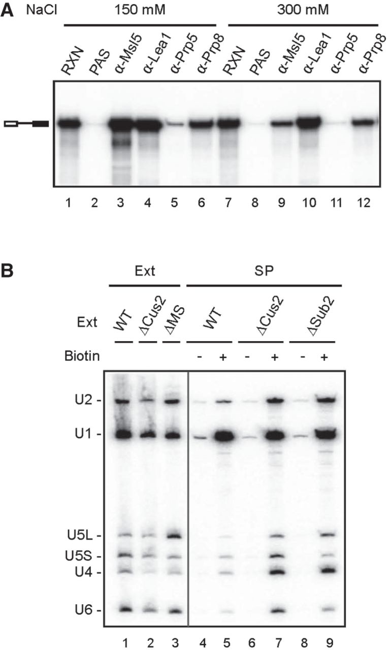
ATP is not essential for the formation of the Pre-B complex in Sub2-depleted extracts. (A) Splicing was performed at 25°C for 15 min in M2M5-supplemented ATP-depleted ΔMS extracts, and then the reaction mixtures were precipitated with no (lanes 2 and 8), anti-Msl5 (lanes 3 and 9), anti-Lea1 (lanes 4 and 10), anti-Prp5 (lanes 5 and 11), or anti-Prp8 (lanes 6 and 12) antibody. The precipitates were washed with standard buffer containing 150 or 300 mM NaCl. RXN, 1/10 of the splicing reaction mixture; PAS, protein A-Sepharose. (B) Northern blot of streptavidin-pulled down spliceosome. Splicing was performed at 25°C for 15 min with pre-mRNA without (lanes 4, 6, and 8) or with (lanes 5, 7, and 9) biotin labels in WT (lanes 4 and 5), ΔCus2 (lanes 6 and 7) or ΔSub2 (lanes 8 and 9) extracts preincubated with glucose for 10 min. The reaction mixtures were precipitated with streptavidin agarose. RNAs from the precipitates and 2 µL of each extract (lanes 1–3) were analyzed by northern blotting using probes for 5 snRNAs. Ext, splicing extract; SP, spliceosome; ΔMS, MUD2, SUB2-double deletion; M2M5, recombinant Mud2-Msl5 dimer.




