


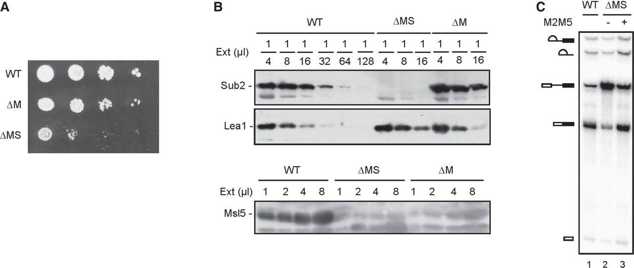
FIGURE 2.
Characterization of ΔMS extracts. (A) Spot assays for growth at 30°C of serial dilutions of WT, ΔM, and ΔMS cells. WT, wild-type; ΔM, MUD2 deletion; ΔMS, MUD2, SUB2-double deletion. (B) Western blot of serial dilutions of WT, ΔM, and ΔMS extracts, probed with anti-Sub2, anti-Lea1, and anti-Msl5 antibodies. WT, wild-type; ΔM, MUD2 deletion; ΔMS, MUD2, SUB2-double deletion; (Ext) Splicing extract. (C) Splicing of ΔMS extracts was performed at 25°C for 30 min with the addition of 2 ng/µL purified recombinant M2M5 proteins. WT, wild-type; ΔMS, MUD2, SUB2-double deletion; M2M5, Mud2-Msl5 dimer.




