


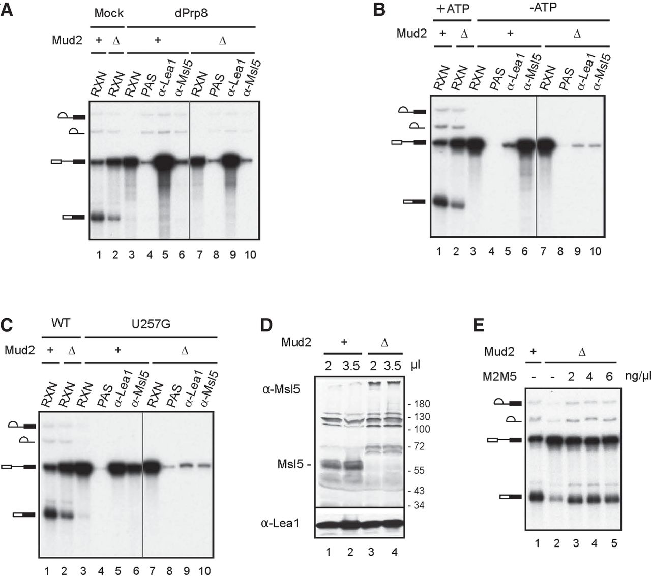
Characterization of ΔMud2 extracts. (A–C) Formation of the prespliceosome (A), the CC (B), and the FIC (C) in ΔMud2 extracts. Splicing was performed at 25°C for 30 min in wild-type (lanes 1 and 3–6) or ΔMud2 (lanes 2 and 7–10) extracts, either depleted of Prp8 (A) or in the absence of ATP (B) using ACT1 pre-mRNA, or in untreated extracts using U257G pre-mRNA (C), followed by precipitation with no (lanes 4 and 8), anti-Lea1 (lanes 5 and 9), or anti-Msl5 (lanes 6 and 10) antibody. (RXN) 1/10 of the splicing reaction mixture; (PAS) protein A-Sepharose; (+) wild-type MUD2; Δ, MUD2 deletion. (D) Western blot of wild-type (lanes 1 and 2) and ΔMud2 (lanes 3 and 4) extracts probed with anti-Msl5 and anti-Lea1 antibodies. (+) Wild-type MUD2; (Δ) MUD2 deletion. (E) Complementation of ΔMud2 extracts with purified recombinant M2M5. Splicing was performed at 25°C for 30 min in wild-type (lane 1) or ΔMud2 (lanes 2–5) extracts, to which various amounts of M2M5 were added. (+) Wild-type MUD2; (Δ) MUD2 deletion; (M2M5) recombinant Mud2-Msl5 dimer.




