


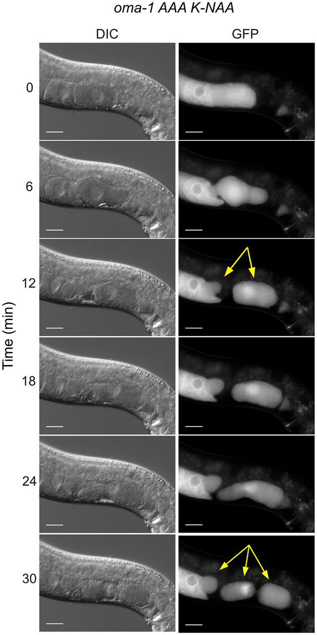
Embryos produced in the oma-1 AAA mutant frequently fragment during ovulation. Still images from a movie of ovulation are shown (Supplemental Movie 1). DIC and GFP stills are shown. The time post-initiation of filming is shown. At the 6 min time point, an oocyte can be seen transitioning the spermatheca, where it is fertilized. By the 12 min time point, the new embryo is fragmented into two pieces, one that remains in the spermatheca, and another in the uterus (yellow arrows). At the 24 min time point, the embryo in the uterus begins to fragment again into two pieces to make a total of three embryo fragments (marked with yellow arrows in the 30 min time point). The scale bars represent 20 µm.




