


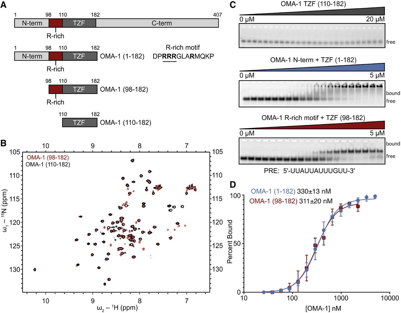
An arginine-rich region in OMA-1 is necessary for RNA-binding activity. (A) Schematic of the OMA-1 protein constructs used in this work. The amino acid regions are noted. The TZF domain and R-rich motif are labeled. (B) HSQC spectrum of OMA-1(92–182) (red) and OMA-1(110–182) (black) showing that both protein variants are folded and adopt similar conformations. (C) Representative EMSA gels with all three MBP-tagged protein variants. Bound and free RNA species are labeled. The RNA sequence used is shown below the gels. (D) Binding curves used to determine the apparent equilibrium dissociation constants (shown) for OMA-1(92–182) (red) and OMA-1(1–182) (blue).




