


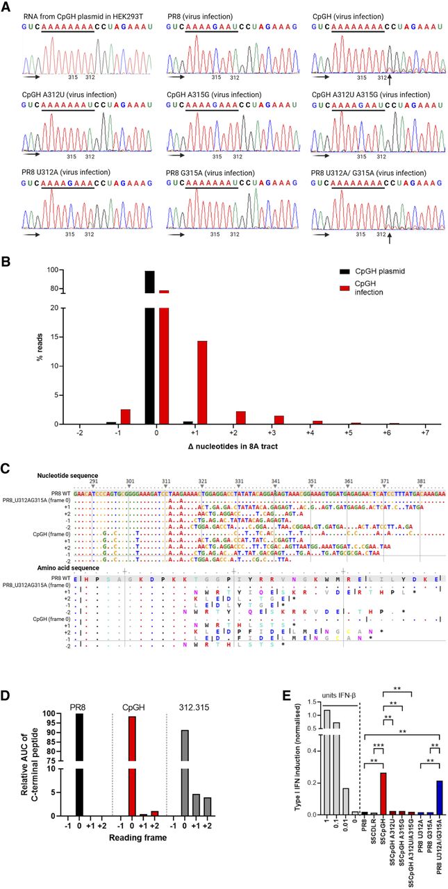
Introduction of the 8A tract into IAV segment 5 resulted in viral polymerase slippage, aberrant protein production, and triggered type I IFN. (A) WT PR8, CpGH, and cognate viruses with switched nucleotides at positions 312 and 315 were used to infect A549 cells at MOI 3 for 8 h, after which RNA was extracted, amplified, and sequenced. Chromatogram traces were examined for evidence of multiple RNA species generated downstream from the 8A sequence of CpGH virus, encoded at nucleotide positions 312–319. Faded black arrows indicate direction of sequencing read. Solid black arrows indicate sites of polymerase slippage. (Top left) CpGH plasmid was transfected into HEK293T cells, and +RNA was produced from a pol.II promoter. (Top middle) WT PR8 virus infection. (Top right) CpGH virus infection. (Middle left) CpGH A312U virus (7A tract) infection. (Middle middle) CpGH A315G virus (4A tract) infection. (Middle right) CpGH A312U A 315G (4A tract) virus infection. (Bottom left) PR8 U312A virus (4A tract) infection. (Bottom middle) PR8 virus G315A (7A tract) infection. (Bottom right) PR8 U312A G315A virus (8A tract) infection. (B) CpGH plasmids and infections were deep sequenced, and percentage of sequence reads with changes in the length of the 8A tract were calculated. (C) Top panel—Nucleotide alignment of PR8, PR8 G312A U315A, and CpGH sequence surrounding the poly(A) site, with alignments to show the nucleotide sequence resulting from +1, +2, −1, and −2 polymerase slippage events. The bottom panel shows the resulting peptide species arising from these transcripts. (D) To examine for the presence of frameshifted peptide production, A549 cells were infected at MOI 3 for 8 h with either WT PR8, CpGH, or PR8 A312U A315G viruses, and NP peptide production was assessed using mass spectrometry to examine for a shift into alternative reading frames. Due to differences in m/z ratios for the peptides unique to +1 and +2 frameshifted translations, relative abundance cannot be compared across peptide species. The −1 frameshift followed by gluC digestion resulted in predicted peptides that were not of sufficient length for detection by mass spectrometry. No peptides predicted to have arisen from the −2 frameshift were detected. (E) Type I interferon competent A549 cells were infected with virus panel for 10 h at MOI 10, after which time supernatant was harvested, UV treated to inactivate infectious virus and assayed for IFN content using HEK Blue cells. HEK Blue cells were also treated with IFN standard (light gray bars). Means of three biological repeats are shown.




