


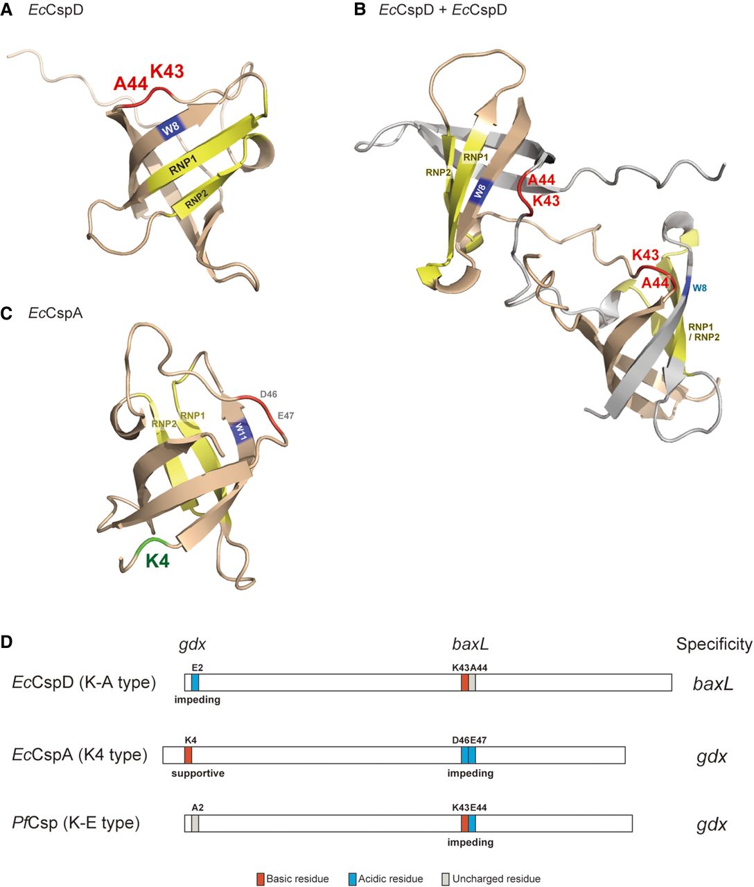
Models of Csp actions and definition of their specificities. (A,B) Predicted structures of EcCspD monomer (A) and homodimer (B). The monomer structure was obtained from AlphaFold Protein Structure Database (Jumper et al. 2021; Varadi et al. 2022). The homodimer structure was predicted with AlphaFold-Multimer (Jumper et al. 2021; Evans et al. 2022); one is shown in wheat, and another is in gray. K43 and A44 in the loop are shown in red. RNA-binding motifs, RNP1 and RNP2, and a tryptophan residue in the vicinity are shown in yellow and blue, respectively. (C) The structure of E. coli CspA monomer (Protein Data Bank ID: 1MJC). K4 in the N-terminal portion is shown in green. (D) Cartoon models summarizing the definition of specificities of the tested Csps. The residues identified in this study are highlighted: acidic residues in blue; basic residues in red; uncharged residues in gray.




