


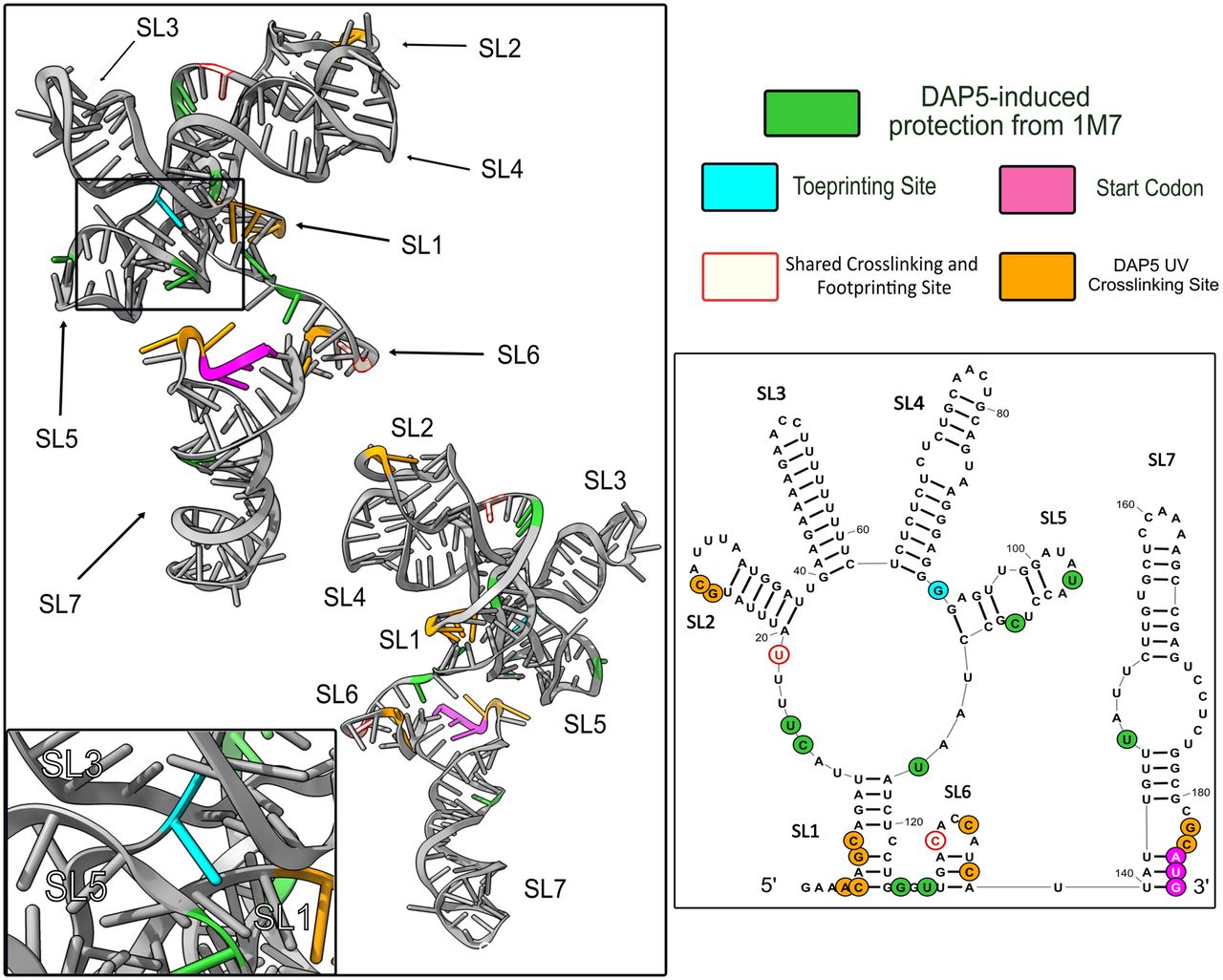
FIGURE 7.
DAP5 contacts mapped on our tertiary model of the FGF-9 5′-UTR mRNA. Tertiary prediction from Figure 6 annotated with DAP5 toeprinting, footprinting, and cross-linking data. Ivory with red outline = RT stops shared between footprinting data (green) and UV cross-linking data (orange). Toeprinting site (nt 93) shown in aqua blue. Start codon (nt 184–186) shown in magenta. All other nucleotides are annotated in black.




