


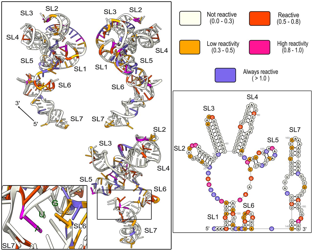
FIGURE 6.
Predicting the FGF-9 5′-UTR tertiary structure. The minimum-free-energy tertiary conformation was predicted by RNAComposer using Figure 2 as a folding constraint. USCF ChimeraX was used to annotate 1M7 reactivity. ΔG = −3060 kcal/mol. SL1–SL5 aggregate closely around the 5′ end of the mRNA, whereas SL6 and SL7 project away. The AUG start codon, tucked between SL5 and SL6, is visualized in an insert (bottom left). SL1 = nt 5−12 and 117–124, SL2 = 20–38, SL3 = 40–62, SL4 = 63–92, SL5 = 94–112, SL6 = 128–138, and SL7 = 140–186. Start codon: 184–186.




