


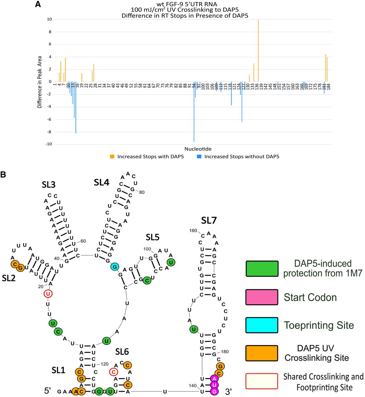
DAP5–FGF-9 5′-UTR UV cross-linking. DAP5 was cross-linked to the wt FGF-9 5′-UTR RNA construct using UV light. For clarity, DAP5 footprinting data are shown alongside UV cross-linking stops. (A) Difference in peak area between RT stops occurring with and without DAP5. (Orange) More frequent stops with DAP5. (Blue) More frequent stops found without DAP5. Size standard peaks (20, 40, 50, 60, 70, 80, 100, 120, 140, 160, 170, and 180 nt) are not shown here. (B) DAP5 footprinting, toeprinting, and UV cross-linking sites mapped on our putative 2D model. Ivory with red outline = RT stops shared between footprinting stops from Figure 4 (green) and UV cross-linking data (orange). Toeprinting site (nt 93) shown in aqua blue. Start codon (nt 184–186) shown in magenta.




