


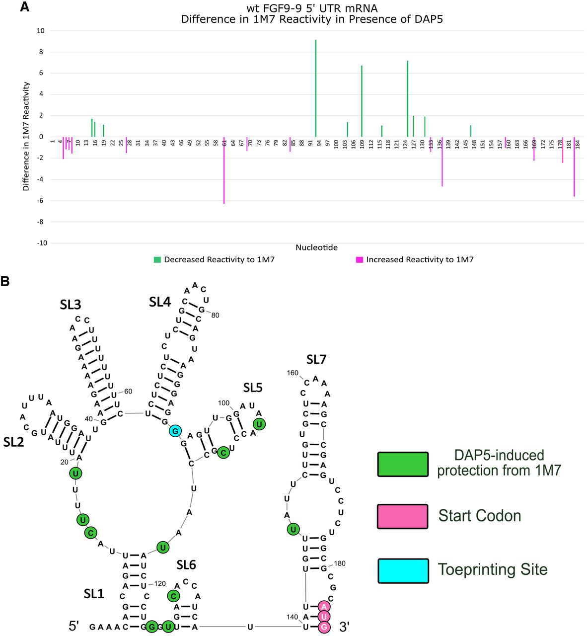
FGF-9 5′ UTR–DAP5 footprinting data. 1M7 reactions were performed on DAP5-bound FGF-9 5′-UTR mRNA. n = 2. (A) Changes in 1M7 reactivity in the presence of DAP5. Normalized 1M7 reactivity in the presence of DAP5 was subtracted from normalized 1M7 reactivity in the absence of DAP5. For clarity, reactivity changes >1 (from a reactive site to a nonreactive site) are shown here. A decrease in 1M7 reactivity > 1 (green) indicates possible contact with DAP5. An increase in 1M7 reactivity (magenta) indicates increased flexibility in the presence of DAP5. Start codon: nt 184–186. Full length of mRNA = 209 nt. Size standard peaks (20, 40, 50, 60, 70, 80, 100, 120, 140, 160, 170, and 180 nt) are not shown here. (B) DAP5-induced protection from 1M7 annotated across the predicted secondary structure of the FGF-9 5′-UTR mRNA. Decreased 1M7 reactivity is annotated in green, with darker green = sharper decrease in reactivity. Toeprinting site (nt 93) from Figure 3 is annotated in aqua blue. AUG (nt 184–186) in magenta.




