


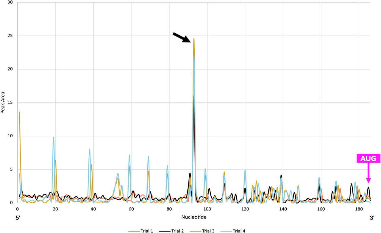
FIGURE 3.
FGF-9 5′ UTR–DAP5 toeprinting data. FGF-9 5′-UTR cDNA was reverse-transcribed from DAP5-bound mRNA. n = 4. Higher peak area corresponds to a higher proportion of RT stops in the presence of DAP5. Size standard peaks visible at 20, 40, 50, 60, 70, 80, 100, 120, 140, 160, 170, and 180 nt. Strong RT stop at nt 93 (black arrow). Start codon: nts184–186 (magenta arrow).




