


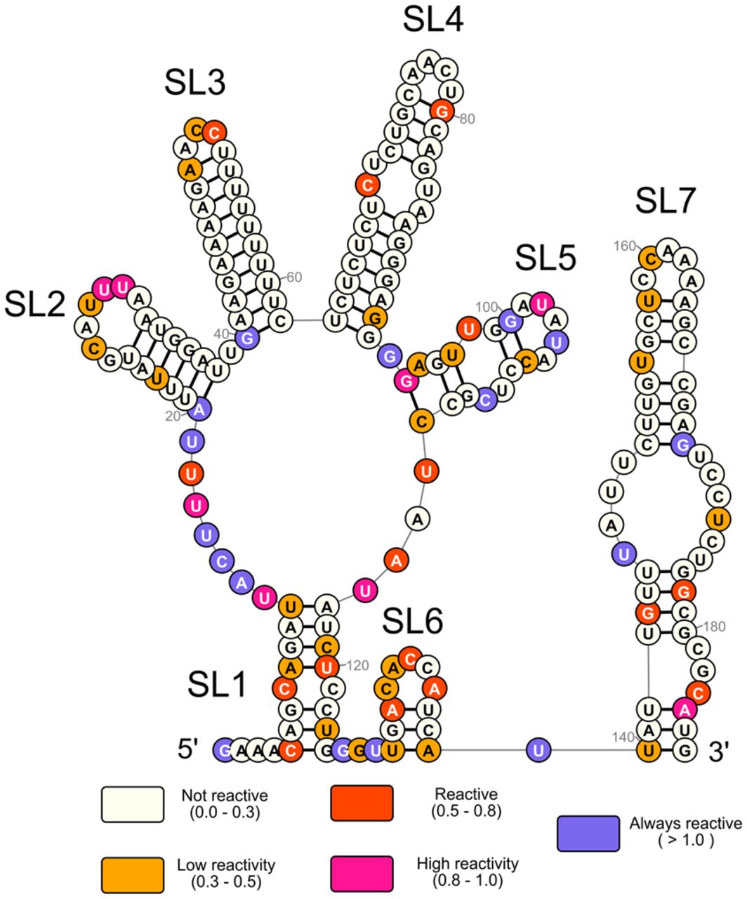
FIGURE 2.
SHAPE-derived 2′ structure of the FGF-9 5′-UTR mRNA. Normalized SHAPE data from three SHAPE reactions were pooled and input into RNAStructure software as a binding constraint. The minimum-free-energy predicted structure (ΔG = −73.1 kJ) was selected. Nucleotides were annotated in RNA2Drawer on a scale from 0 (ivory, unreactive to 1M7) to 1.0 and above (medium slate blue, always reactive to 1M7, seven SL were predicted to form). The AUG start codon is visible at the base of SL7.




