


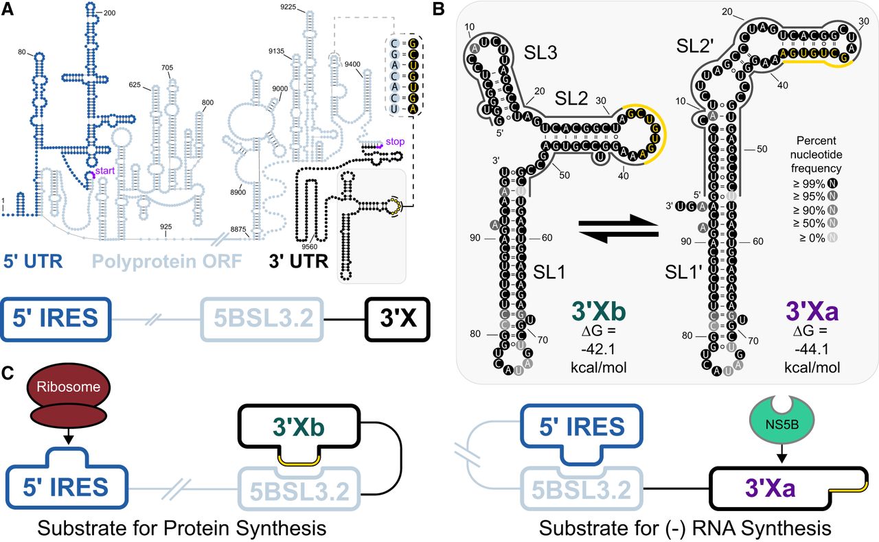
Architecture of the hepatitis C virus (HCV) genome. (A) The HCV genome is highly structured with extensive intragenomic base-pairing interactions and can be partitioned into three regions: the 5′ UTR (nt: 1–341), the ORF (nt: 342–9442), and the 3′ UTR (nt: 9443–9678). Figure adapted from Wan et al. (2022). (B) The 3′ UTR is terminated by a 98 nt sequence known as 3′X. It is highly conserved, with the color of each nucleotide representing the frequency with which it appears in the NCBI GenBank. There are two different experimentally supported secondary structures for this RNA (3′Xb and 3′Xa). The major structural differences between the two conformations are largely confined to the first 55 nt associated with either stem–loops SL2/3 or SL2′. Consequently, the two conformations either present or sequester the 7 nt associated with the k-sequence (yellow), which can participate in a distal interaction with 5BSL3.2 in the ORF (A, inset). (C) The presence of this k-5BSL3.2 interaction (left) leaves the IRES free to promote viral protein synthesis. Alternatively, 5BSL3.2 can also interact with the IRES (right), which leaves 3′X free to adopt a conformation (3′Xa) that favors RNA synthesis.




