


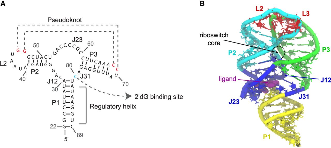
FIGURE 1.
(A,B) The secondary structure and X-ray crystallographic structure of 2′-dG riboswitch used for explicit-solvent simulation. (Yellow) P1 helix, (cyan) P2 helix and the backbone of loop L2, (green) P3 helices and the backbone of loop L3, (red) loops L2 and L3 which form an HHH-type PK, (blue) three-way junction, which contributes major interactions with the ligand 2′-dG, (purple) 2′-dG molecule shown in van der Waals representation.




