


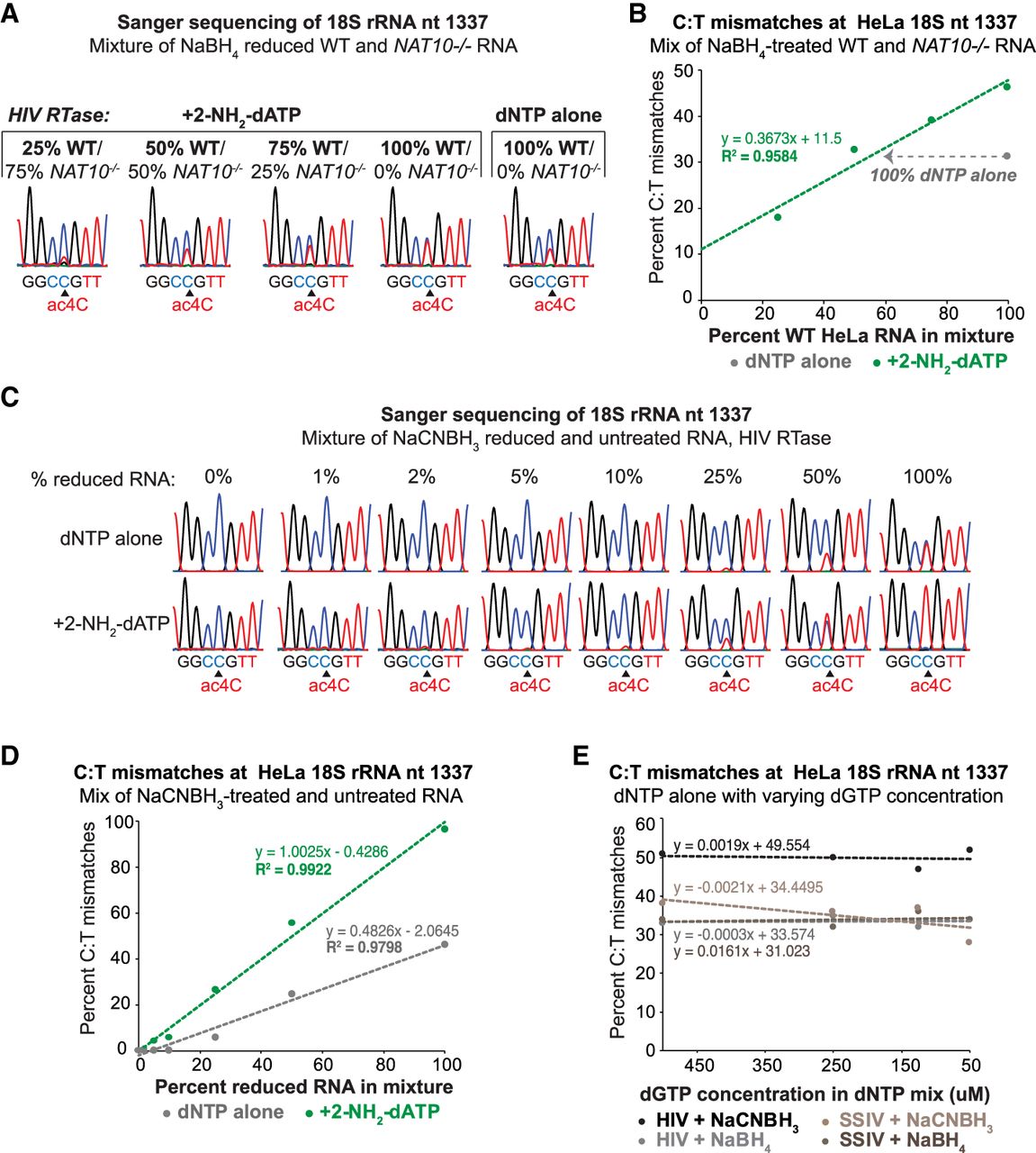
Quantitative detection of ac4C with 2-amino-dATP in Sanger sequencing. (A) NaBH4 reduction of total RNA from wild-type (WT) and NAT10−/− HeLa cells mixed at the indicated ratios and cDNA synthesis with HIV RTase and the canonical dNTPs (100% dGTP) or with dGTP partially substituted with 2-amino-dATP (75% analog:25% dGTP). Sanger sequencing results of a PCR amplicon surrounding the ac4C site in 18S rRNA helix 34. (B) Quantification of C:T mismatch levels through Sanger chromatogram peak heights at 18S rRNA nt 1337 from A. The trendline depicts a linear increase in C:T mismatch detection with increasing percent WT RNA. The gray dotted arrow indicates the C:T mismatch level in parallel processed samples from 100% WT RNA reverse transcribed with dNTP alone. (C) Sanger sequencing traces of RT-PCR amplicons surrounding the helix 34 ac4C site from mixtures of untreated and NaCNBH3-treated HeLa RNA, as indicated. RT was performed with HIV RTase and the canonical dNTPs (100% dGTP), or with dGTP partially substituted with 2-amino-dATP (75% analog:25% dGTP). (D) Quantification of C:T mismatch levels through Sanger chromatogram peak heights at 18S rRNA nt 1337 from C. The trendlines depict enhanced mismatch detection in reactions including 2-amino-dATP. (E) Quantification of C:T mismatch levels at 18S rRNA nt 1337 through Sanger chromatogram peak heights from NaBH4 and NaCNBH3-reduced WT HeLa RNA. cDNA synthesis was accomplished with HIV and SSIV RTases with decreasing concentrations of dGTP starting from the 500 µM used in canonical dNTP mixtures.




