


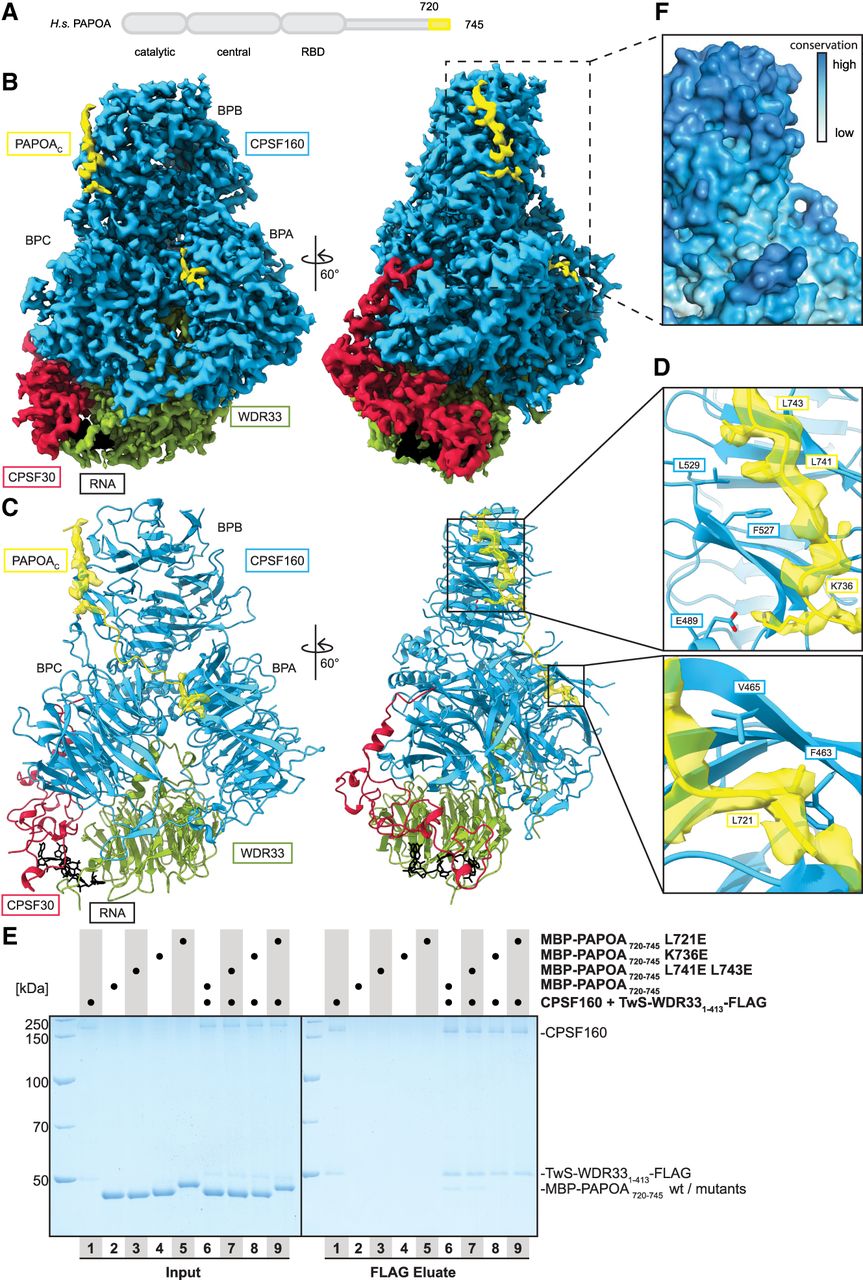
PAPOAC binds the mPSF subunit CPSF160. (A) Domain organization of human PAPOA. The truncated construct used here is indicated in yellow. (B) Segmented single-particle cryo-EM reconstruction of mPSF-PAPOAC at a global resolution of 2.79 Å shown in two different orientations. The densities for CPSF160, WDR33, CPSF30, PAPOAC, and RNA are colored in blue, green, red, yellow, and black, respectively. The CPSF160 β-propeller domains are indicated. (C) Structural model of mPSF overlay with the computationally predicted model of PAPOAC with the segmented cryo-EM density of PAPOAC displayed in transparent yellow, shown in the same orientations and colors as in B. (D) Close-up of the cryo-EM density of the binding interfaces of CPSF160–PAPOAC. The refined model is shown and the segmented cryo-EM density of PAPOAC is displayed in transparent yellow. Labeled PAPOAC residues were changed to glutamic acid. (E) Coomassie-stained SDS-PAGE analysis of pull-down experiment showing that the negatively charged mutations in the CPSF160–PAPOAC interface impair or disrupt the interaction. (F) Close-up of the binding site of CPSF160 for PAPOAC shown in surface representation with residues colored by sequence conservation, from low conservation in white to high conservation in blue.




