


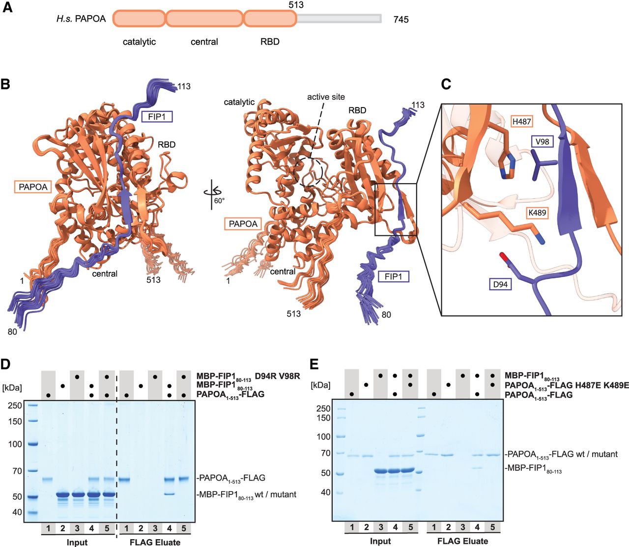
Identification of the PAPOA-binding region of FIP1. (A) Domain organization of human PAPOA. The truncated construct used here is indicated in orange. (B) Overlay of 25 computationally predicted models of the PAPOA1–513–FIP180–113 complex, shown in two orientations. The PAPOA structured domains and active sites are indicated. (C) Close-up of the putative binding interface. Labeled residues were changed to arginine (FIP1) or glutamic acid (PAPOA). (D) Coomassie-stained SDS-PAGE analysis of pull-down experiment showing that positively charged FIP1 mutations in the predicted PAPOA–FIP1 interface disrupt the interaction. (E) Coomassie-stained SDS-PAGE analysis of pull-down experiment showing that negatively charged PAPOA mutations in the predicted PAPOA–FIP1 interface disrupt the interaction.




