


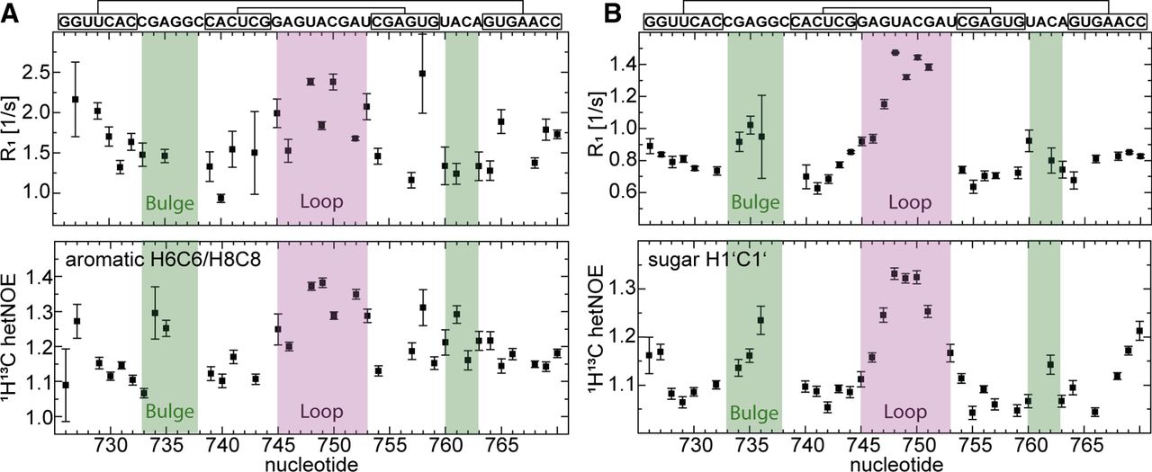
FIGURE 7.
1H, 13C R1 rates (upper panels), and heteronuclear NOE of s2m Delta (lower panels). For all measurements, separate experiments were recorded for (A) aromatic C6H6/C8H8 and (B) ribose C1′H1′ resonances. The secondary structure of the RNA as determined by NMR is given at the top. The loop region (pink) and internal loop region (green) are highlighted in the plots. The notation of nucleotides corresponds to the position in the genome −29,000.




