


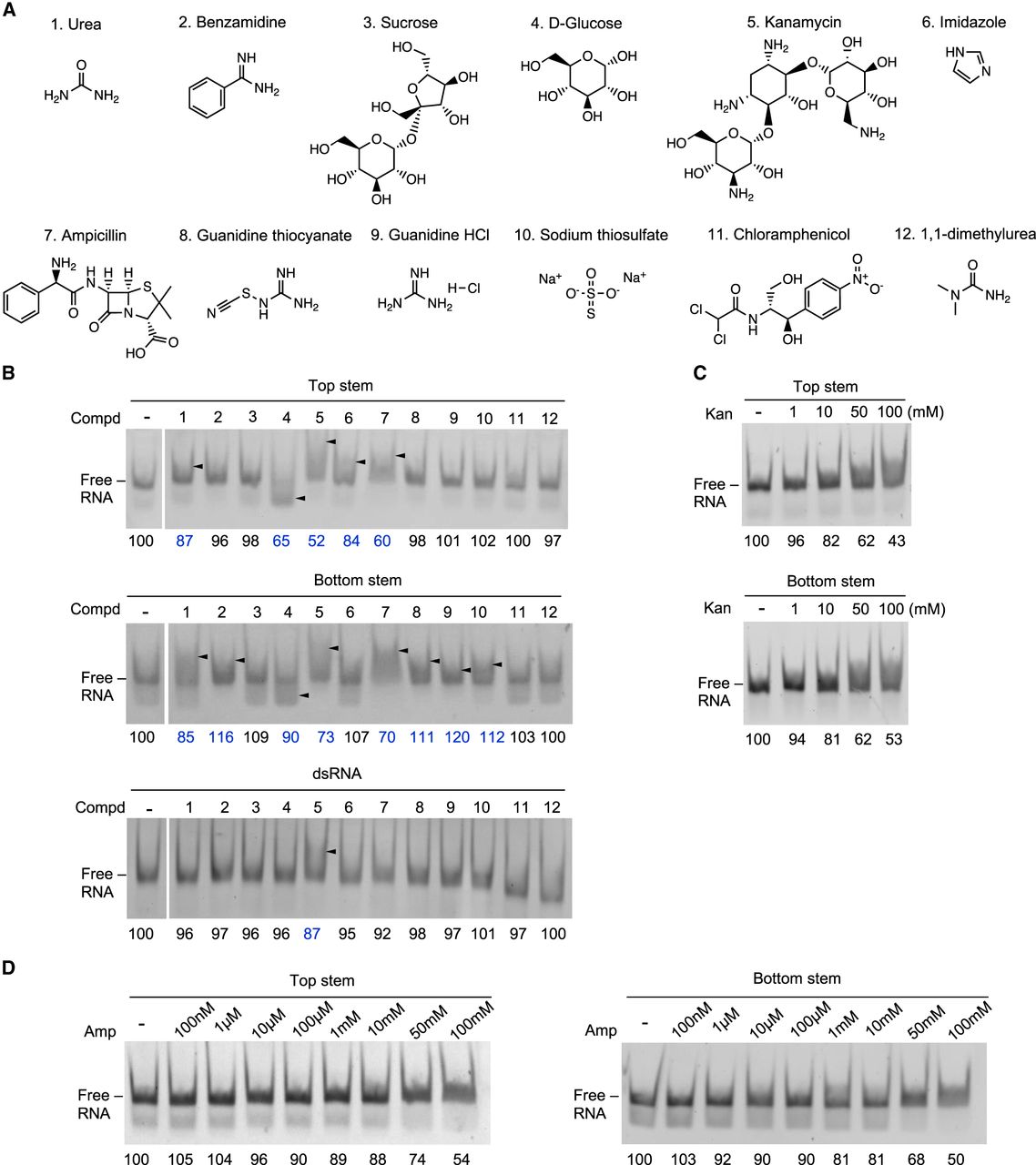
Screening for the RNA-binding potential of selected chemicals. (A) Twelve chemicals assayed for their ability to bind the isolated ZIKV SLA stem constructs. (B) Gel shift assay. The chemicals listed in A (50 mM) were incubated with top and bottom stem RNA and the negative control dsRNA (150 nM), and the mixtures were resolved via native PAGE. Band shifts are indicated by a triangle arrow to the right side of the lane. Band intensities of the remaining RNA were quantified and normalized against the control lane. Interaction between the chemical and RNA was identified via deviation of the band intensity by more than 10% from that of the control lane. (C) Gel shift assay with kanamycin. Top and bottom stem RNA (1 µM) was incubated with an increasing concentration of kanamycin from 1 to 100 mM. Quantified band intensities for dsRNA are shown below the gel. (D) Gel shift assay with ampicillin. Top and bottom stem RNA (1 µM) was incubated with an increasing concentration of ampicillin from 100 nM to 100 mM.




