


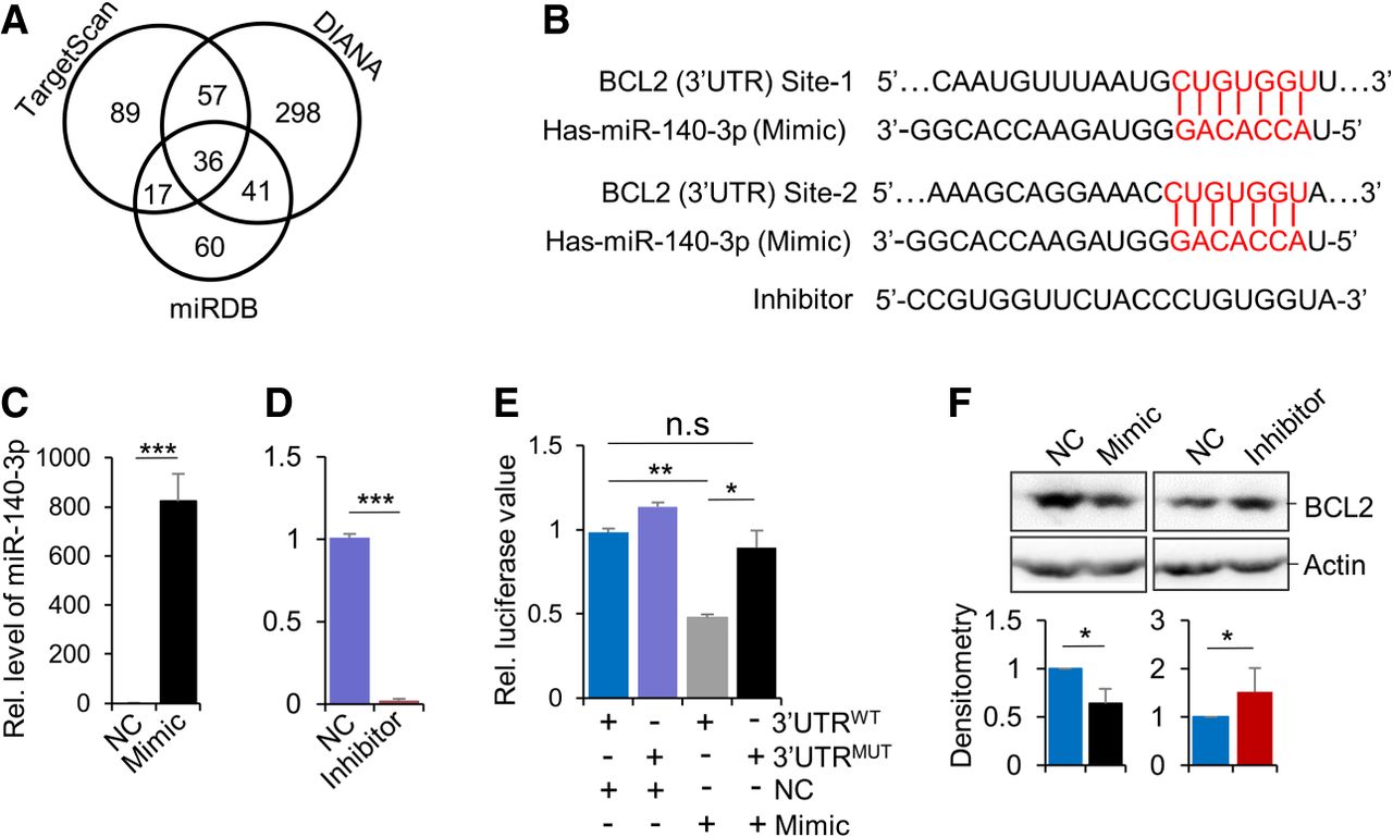
Identification of BCL2 as a direct target of miR-140-3p. (A) Venn diagram of the miR-140-3p target genes from TargetScan, miRDB, and DIANA databases. (B) Predicted binding sites (in red) of miR-140-3p in the 3′ UTRs of BCL2. The full-length sequence of miR-140-3p (Mimic) and inhibitor was presented. (C,D) The miR-140-3p mimic (C), inhibitor (D), and NC were transfected into MOLM-13 cells, respectively. Expression of miR-140-3p was measured by RT-qPCR and quantified from three biological repeats. The sequence information of synthesized miR-140-3p mimic, inhibitor, and respective NC is provided in Supplemental Table S3. (E) The psiCHECK-2 dual-luciferase reporter plasmid harboring wild-type BCL2 3′ UTR (WT) or mutated version (both of the two binding sites were replaced with 5′-GACACCA-3′), together with miR-140-3p Mimics and NC were transfected into 293T cells, respectively. The effects of BCL2 3′-UTR WT, its mutant, and miR-140-3p Mimics on luciferase intensity were evaluated. The quantification was performed using data from two biological replicates. (F) The miR-140-3p Mimic, inhibitor, and respective NC were transfected into MOLM-13 cells, respectively. Indicated proteins were detected by western blotting. (Bottom) Quantification from three biological repeats. Data represent the mean + SD (C–F). (*) P < 0.05, (**) P < 0.01, and (***) P < 0.001, by Student's t-test (C,D,F) and one-factor ANOVA with a post hoc t-test (E).




