


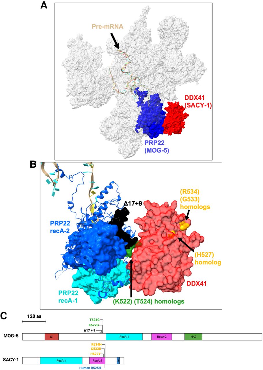
Diagram of sacy-1 and mog-5 mutations and the locations of their homologs in C*. (A) Model of C* structure based on coordinates from Dybkov et al. (2023), PDB ID 8C6J, image generated with ChimeraX (Pettersen et al. 2021). Shown are the pre-mRNA 3′ lariat intermediate (tan), PRP22 (blue), and DDX41 (red), and all other spliceosomal components are in transparent gray. (B) Closer view of PRP22/DDX41 interface showing the positions in human that correspond to C. elegans mutations, the same orientation as Figure 5A. Shown are homologs of SACY-1 (H527), (G533), and (R534) (yellow), MOG-5(K522, T524) (green), the region of amino acids that are altered in mog-5(Δ17 + 9) (black), the RecA-1 domain of PRP22 (light teal), and the RecA-2 domain of PRP22 (purple). (C) Protein structure diagram of SACY-1 and MOG-5. Scale bar shows the distance measured in amino acids for both proteins. Portions of amino acid sequences corresponding to domains are colored. Domain boundaries were taken from SMART (Letunic et al. 2021). Z stands for ZnF C2HC. Mutations in C. elegans are shown above the protein diagram, whereas a mutation in human is shown below.




