


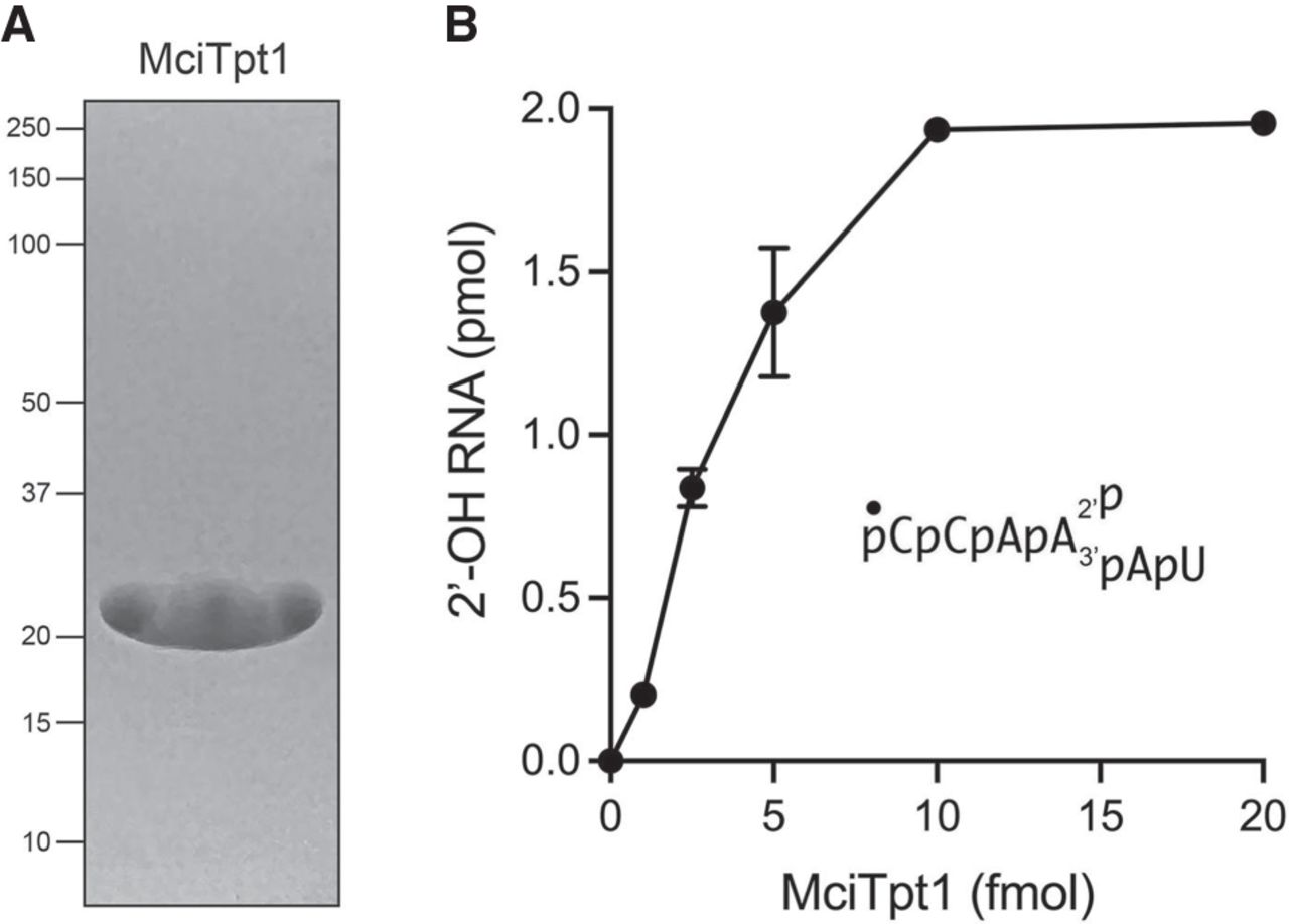
2′-phosphotransferase activity of recombinant Mucor Tpt1. (A) An aliquot (7.5 µg) of the recombinant MciTpt1 preparation was analyzed by SDS-PAGE. The Coomassie blue-stained gel is shown. The positions and sizes (kDa) of marker proteins are indicated on the left. The expected molecular weight of MciTpt1 is 24 kDa. (B) Reaction mixtures (10 µL) containing 100 mM Tris-HCl, pH 7.5, 1 mM NAD+, 0.2 µM (2 pmol) 5′ 32P-labeled 2′-PO4 branchpoint-containing 6-mer RNA oligonucleotide (as shown), and 0, 1, 2.5, 5, 10, or 20 pmol MciTpt1 were incubated at 37°C for 30 min. The reactions were quenched by the addition of three volumes of cold 90% formamide, 50 mM EDTA. The samples were analyzed by electrophoresis (at 55 W constant power) through a 40-cm 20% polyacrylamide gel containing 7 M urea in 45 mM Tris-borate, 1 mM EDTA. The 5′-radiolabeled 2′-PO4 substrate RNA and the 2′-OH product RNA were visualized by scanning the gel with a Fujifilm FLA-7000 imaging device. The extents of product formation were quantified by analysis of the gel scans in ImageQuant and are plotted as a function of input MciTpt1. Each datum is the average of four independent titration experiments ± SEM.




