


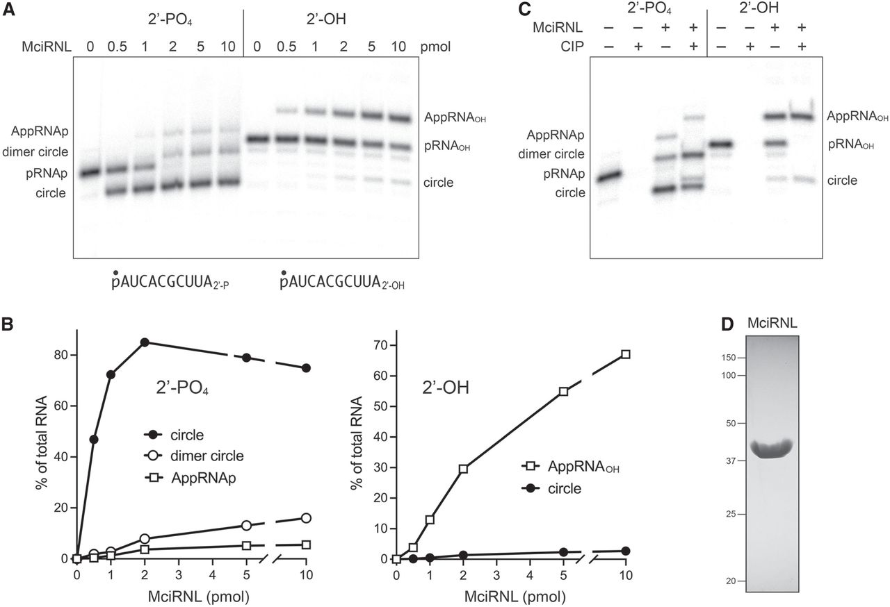
RNA ligase activity of recombinant MciRNL. (A) Reaction mixtures (10 µL) containing 50 mM Tris-acetate, pH 6.5, 5 mM MgCl2, 0.1 µM (1 pmol) 5′ 32P-labeled 10-mer RNA with either a 3′-OH,2′-PO4 end (left side) or a 3′-OH,2′-OH end (right side) and increasing amounts of MciRNL as specified were incubated at 37°C for 5 min. The reactions were quenched with an equal volume of 95% formamide/50 mM EDTA, and the products were analyzed by electrophoresis (at 58 W constant power) through a 40-cm 20% polyacrylamide gel containing 7 M urea in 45 mM Tris-borate, 1 mM EDTA. The 5′-radiolabeled RNAs were visualized by scanning the gel with a Fujifilm FLA-7000 imaging device. The positions and identities of the RNA substrates, RNA-adenylate intermediates, and ligated products are indicated on the left and right. (B) The distributions of radiolabeled RNAs in each lane in panel A were quantified by analysis of the gel scans in ImageQuant and are plotted as a function of input MciRNL for the ligation reactions with the 2′-PO4 (left panel) and 2′-OH (right panel) RNA substrates. (C) Product analysis via treatment with CIP (calf intestine alkaline phosphatase). Ligation reactions containing 1 pmol 5′ 32P-labeled pRNA2′p and 10 pmol MciRNL (where indicated by +) were incubated at 37°C for 5 min. The mixtures were then incubated for 10 min at 37°C with 5 U CIP (purchased from NEB) where indicated by + above the lanes. The mixtures were quenched and then analyzed by urea-PAGE as described in A. (D) An aliquot (10 µg) of the recombinant MciRNL preparation was analyzed by SDS-PAGE. The Coomassie blue-stained gel is shown. The positions and sizes (kDa) of marker proteins are indicated on the left. The expected molecular weight of MciRNL is 43 kDa.




