


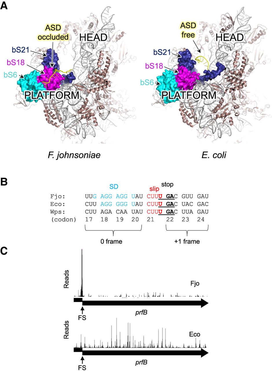
Flavobacterium johnsoniae and Escherichia coli ribosomes differ in structure and function. (A) Comparison of the 30S subunit from F. johnsoniae (left) and E. coli (right), viewed from “above” the platform domain. Proteins bS21, bS18, and bS6 form a pocket that binds the 3′ tail and occludes the ASD in F. johnsoniae. (B) Sequence comparison of the prfB frameshift site in F. johnsoniae (Fjo), E. coli (Eco), and Winogradskyella psychrotolerans (Wps). Codon numbers (based on F. johnsoniae) are indicated below, with the zero and +1 reading frames indicated. The SD, slippery sequence (slip), and UGA stop codon (stop) are shown in blue, red, and bold-underscore, as indicated. (C) Ribosome distribution along the prfB coding region in F. johnsoniae (Fjo) and E. coli (Eco). Reads (arbitrary scale) were mapped with respect to the central nucleotide of the predicted P codon. (FS) Frameshift site.




