


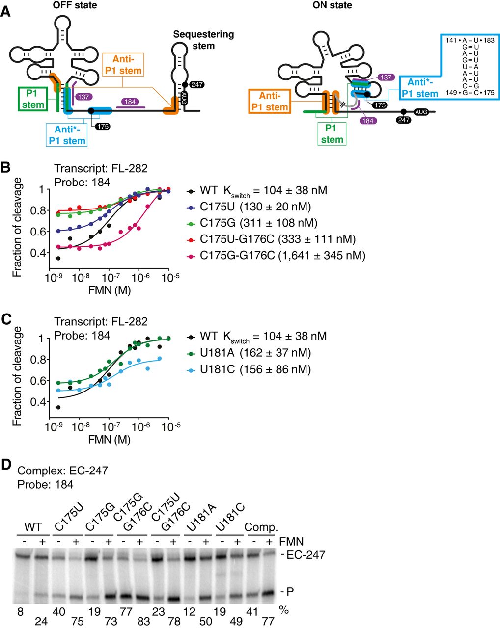
The ribB riboswitch contains an additional helical domain in the FMN-free ON state. (A) Schematics representing the ribB riboswitch in the FMN-bound OFF and FMN-free ON conformations. The regions involved in the predicted P1, anti*-P1, and anti-P1 stems are shown in green, blue, and orange, respectively. While the P1 stem is formed in the OFF state, the anti*-P1, and anti-P1 stems are formed in the ON state. The structure of anti*-P1 stem is shown on the right. The location of the RNase H probes and pause sites are indicated in purple and black, respectively. The line in gray in the ON state represents a section for which no attempt was made to draw the secondary structure. (B,C) Kswitch determination using the probe 184 for the FL transcripts. The mutants performed at positions C175/G176 (B) and U181 (C) were analyzed. Cleavage assays were performed using a range of 2 nM to 10 µM FMN. The fitting analyses were performed by normalizing the cleavage fraction to 1, and the Kswitch values are indicated for each mutant. The experiments were performed in duplicate, and the most representative data are shown. (D) RNase H probing assays obtained on EC-247 ribB nascent transcripts with the probe 184. Experiments were performed without (−) and with (+) 100 µM FMN. The WT and the mutants used are indicated at the top. The FL EC-247 transcripts (EC-247) and cleaved products (P) are indicated on the right of the gel. The experiments were performed in triplicate, and the most representative data are shown.




