


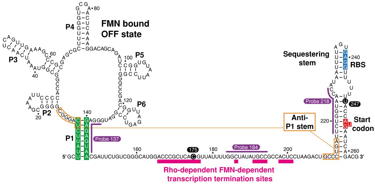
The sequence and secondary structure of the ribB riboswitch of E. coli. The nucleotide numbering is based on previous studies (Winkler et al. 2002b; Hollands et al. 2012). While the structure of the aptamer is depicted according to the consensus structure (Vitreschak et al. 2002; Winkler et al. 2002b), the organization of the expression platform is based on previous analyses (Hollands et al. 2012). The structure is depicted as when bound to FMN (OFF state). The aptamer domain consists of stems P1–P6, and the expression platform encompasses the sequestering stem. The P1 stem is highlighted in green. The anti-P1 stem that is forming in the FMN-free ON state is shown in orange. The ribosome-binding site (RBS) and AUG start codon are shown in blue and red, respectively. The pause sites 175 and 247 are indicated in black circles. The RNase H probes 137, 184, and 218 are shown in purple. Rho-dependent transcription termination sites are shown in pink (Hollands et al. 2012).




