


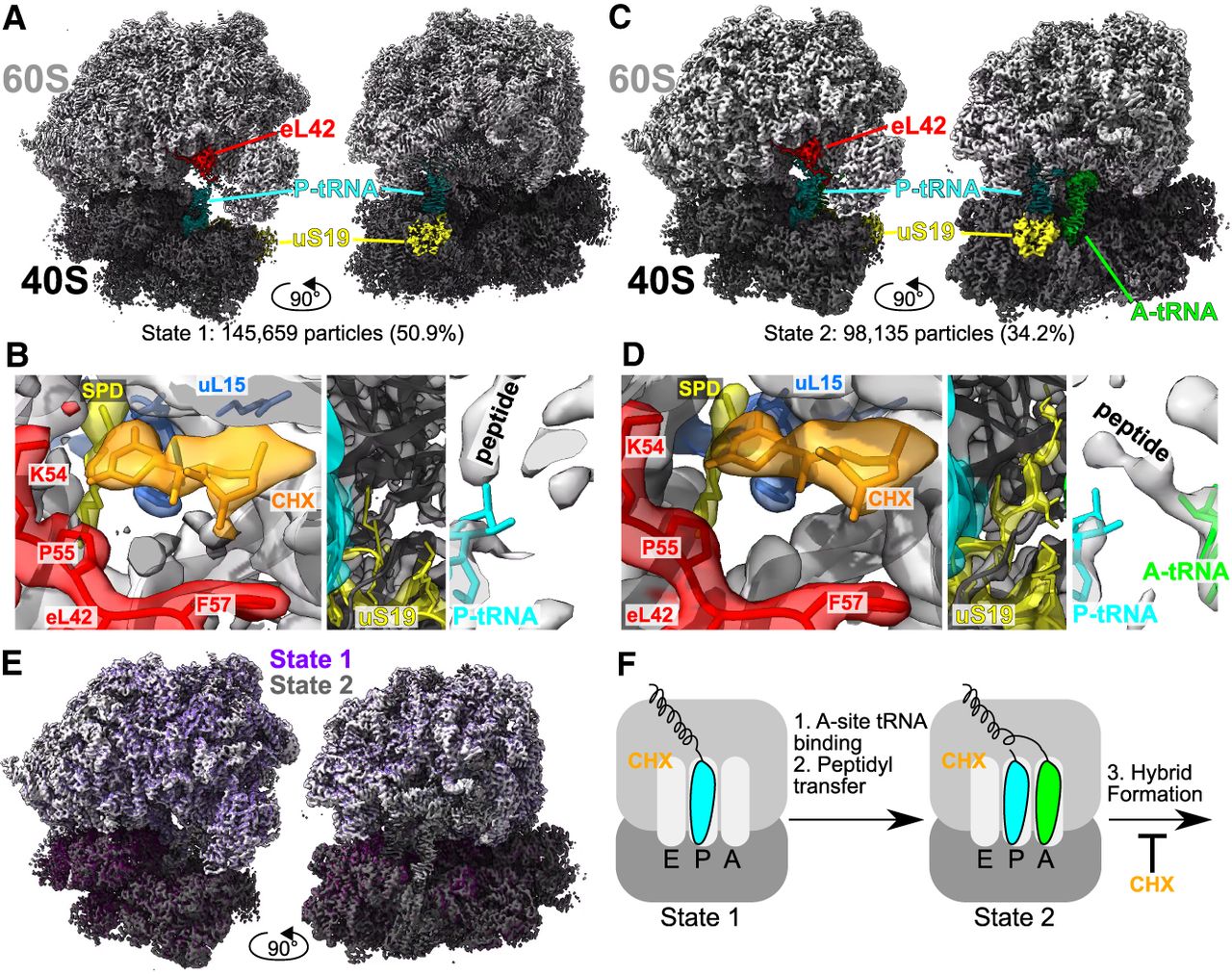
Cycloheximide stabilizes classical PRE ribosomes in C. elegans. (A) Single-particle cryoEM reconstructed map of State 1, with indicated features highlighted; large subunit in light gray and small subunit in dark gray. (B) Highlight of indicated ribosome features. (Left) Cycloheximide (CHX, orange) and spermidine (SPD, yellow) bound near the E-site at eL42 (red) and uL15 (blue). (Middle) uS19 C-terminal tail (yellow) in between the P- and A-sites. (Right) Peptidyl transferase center with 3′OH of P-tRNA in blue and path of the nascent peptide indicated. (C) Single-particle cryoEM reconstructed map of State 2, with indicated features highlighted. (D) As in B, but for State 2. (E) Overlay of State 1 (purple) and State 2 (gray), showing high similarity throughout the map. State 1 large subunit in light purple, and small subunit in dark purple. (F) Model of ribosomal states obtained via single-particle cryo-EM. CHX, cycloheximide. P-site tRNA is in cyan, A-site tRNA is in green, and the peptide is attached as indicated.




