


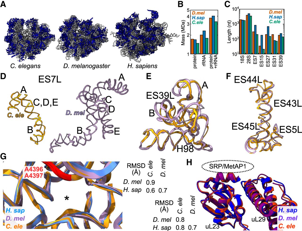
The C. elegans ribosome is streamlined but conserved. (A) Ribosomes from the indicated organisms are shown, with ribosomal proteins in blue and ribosome RNA in gray. D. mel and human structures from Anger et al. (2013), with eEF2 removed. (B) Sizes of the protein, rRNA, and both components of the ribosome in the respective organisms. We included masses for eL28, RACK-1, and the stalk proteins in C. elegans. For rRNA, we included masses for the expansion segments, regardless of whether they were present in the respective models. Similarly, we report the mass for full-length ribosomal proteins, regardless of the presence or absence of N- and C-termini in the maps. (C) Lengths in nucleotides of the indicated rRNAs or 28S expansion segments. As in B, we report the length of the entire expansion segment, not just the segments in the respective models. (D) ES7L from C. elegans (orange) and D. melanogaster (purple). The A, B, C, D, and E arms of ES7L are indicated. (E) ES39L from C. elegans (orange) and D. melanogaster (purple), aligned in three-dimensional space, with A and B arms and Helix 98 indicated. (F) Same as E for ES5L and ES43-45L. (G) Alignment of 28S rRNA within 20 Å of the peptidyl transfer center of the human (blue), Drosophila (purple), and C. elegans (orange) ribosomes. Asterisk indicates the location of the 3′ end of the P-site tRNA. Two universally conserved adenosines (human A4396 and A4397) are highlighted in red. RMSD table measuring 3D similarity for the PTC from the indicated species shown at right. (H) 3D alignment of uL23 and uL29, with the binding site of protein maturation and secretion machineries shown. Color scheme and RMSD table as in G.




