


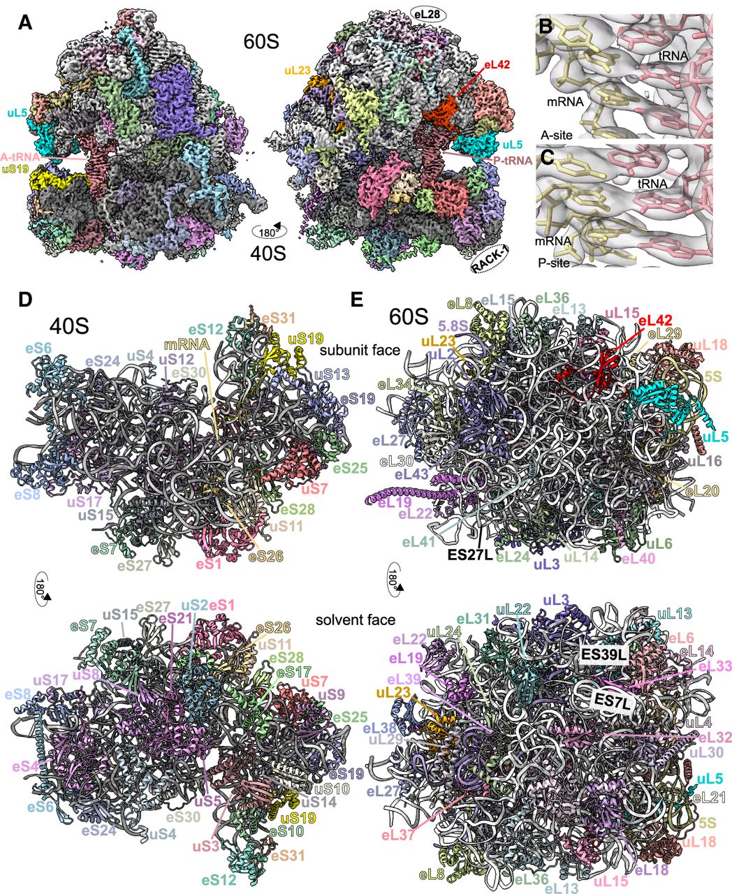
High-resolution structure of a C. elegans ribosome. (A) Segmentation of the reconstructed map of the C. elegans ribosome. 28S rRNA in white, 18S rRNA in gray, A- and P-site tRNAs in pink, and ribosomal proteins are colored. Highlighted in brighter colors and proteins noted: uS19 (yellow), uL5 (blue), uL23 (orange), eL42 (red). RACK-1 and eL28 were not observed, but their approximate locations are indicated. Panels B and C show the zoom of map (light gray) and model (mRNA in yellow and tRNA in pink) for the A- and P-sites, respectively. Note that map heterogeneity is expected in the A- and P-sites as ribosomes were prepared from whole C. elegans and would be expected to contain a diversity of sequences. (D) Atomic model of the C. elegans 40S, with coloring as in A and ribosomal features indicated. Top panel shows the 60S-facing side, with mRNA in light yellow. Bottom panel shows the solvent-facing side. tRNAs hidden for clarity. (E) Atomic model of the C. elegans 60S, with coloring as in A. Top and bottom panels show 40S- and solvent-facing sides, respectively. Expansion segments (ES) 27L, 7L, and 39L are highlighted. tRNAs were omitted from (D) and (E).




