


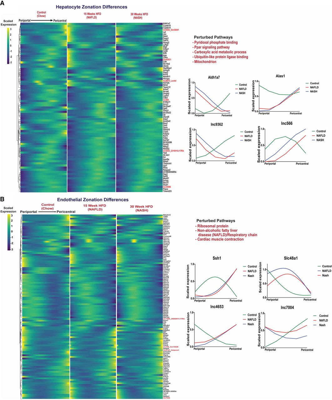
FIGURE 5.
Perturbation of zonation in NAFLD and NASH liver. Matched heatmaps of genes (PCGs and lncRNAs) that are differentially zonated between control, NAFLD, and NASH livers at FDR < 0.001, in hepatocytes (A), and in endothelial cells (B). Pathways perturbed were identified by DAVID functional enrichment analysis of the differential PCGs (Supplemental Table S4I,J). Shown at the right are zonation profiles for select genes for each cell type across three conditions (chow diet, NAFLD, NASH).




