


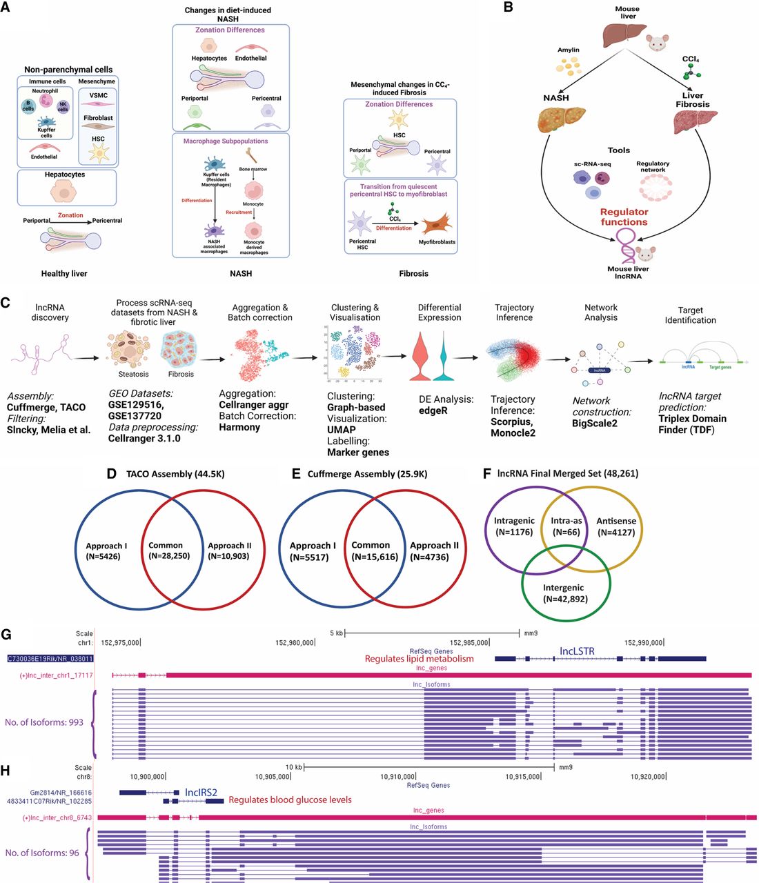
LncRNA discovery and role in NASH and liver fibrosis analyzed using single-cell technology. (A) Healthy liver is comprised of hepatocytes and nonparenchymal cells, notably endothelial cells, mesenchymal cells, and immune cell populations. With the emergence of NASH, changes in gene expression and zonation occur in hepatocytes and endothelial cells and new macrophage subpopulations emerge. In CCl4-induced liver fibrosis, changes in hepatic mesenchymal cells include zonation differences in hepatic stellate cells (HSC) and the transition of pericentral stellate cells to collagen-producing myofibroblasts. (B) Discovery of regulatory roles of lncRNAs in two liver disease models: Amylin diet-induced NASH and CCl4-induced liver fibrosis. (C) Computational workflow for characterization of functional roles of liver-expressed lncRNAs used in this study. (D,E) Numbers of mouse (mm9) liver lncRNAs discovered using TACO assembly (D) and Cuffmerge assembly (E), with two different filtering approaches, I and II. (F) Final set of 48,261 mouse lncRNAs, classified based on their location with respect to PCGs after conversion to mouse mm10 genomic coordinates. (G,H) Two liver-expressed lncRNAs, both found to be comprised of many novel isoforms, a subset of which is shown.




