


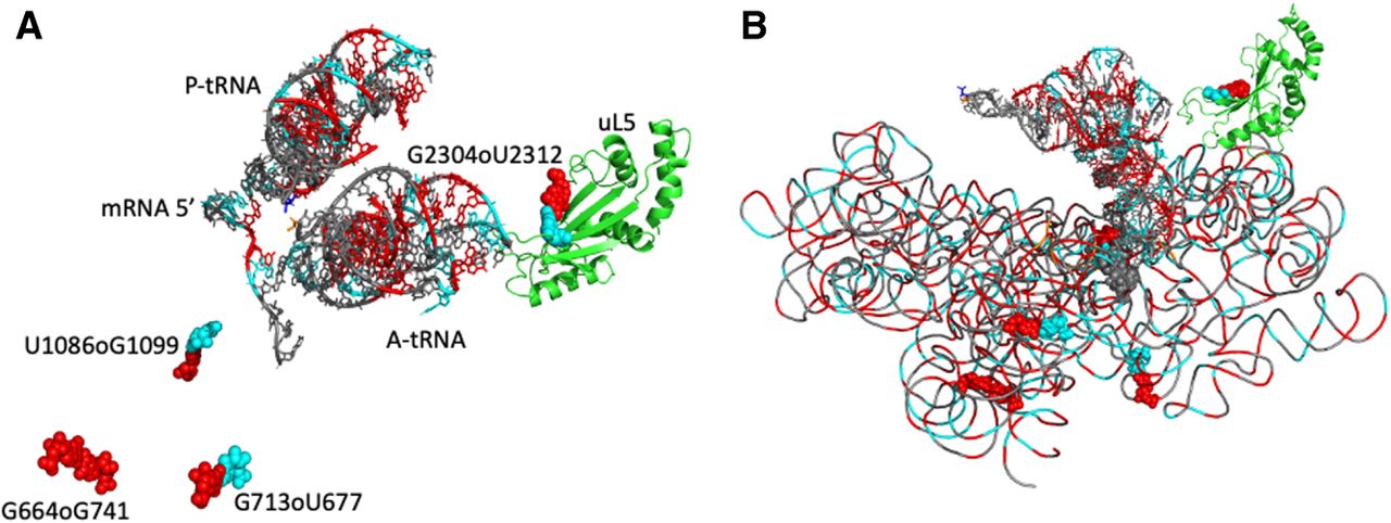
FIGURE 8.
Two views of the molecular environment around the anionic pairs and their proximity within the ribosome structure. The guanines are colored red and shown as van der Waals spheres with the uracils colored in cyan. (A) The A-tRNA and P-tRNA bound to the mRNA (with van der Waals spheres at the right) are shown. The protein uL5 in the 23S rRNA is colored green. (B) The backbone of the whole 16S rRNA is superimposed (with the same color code). The drawings are from 7K00 (Watson et al. 2020).




