


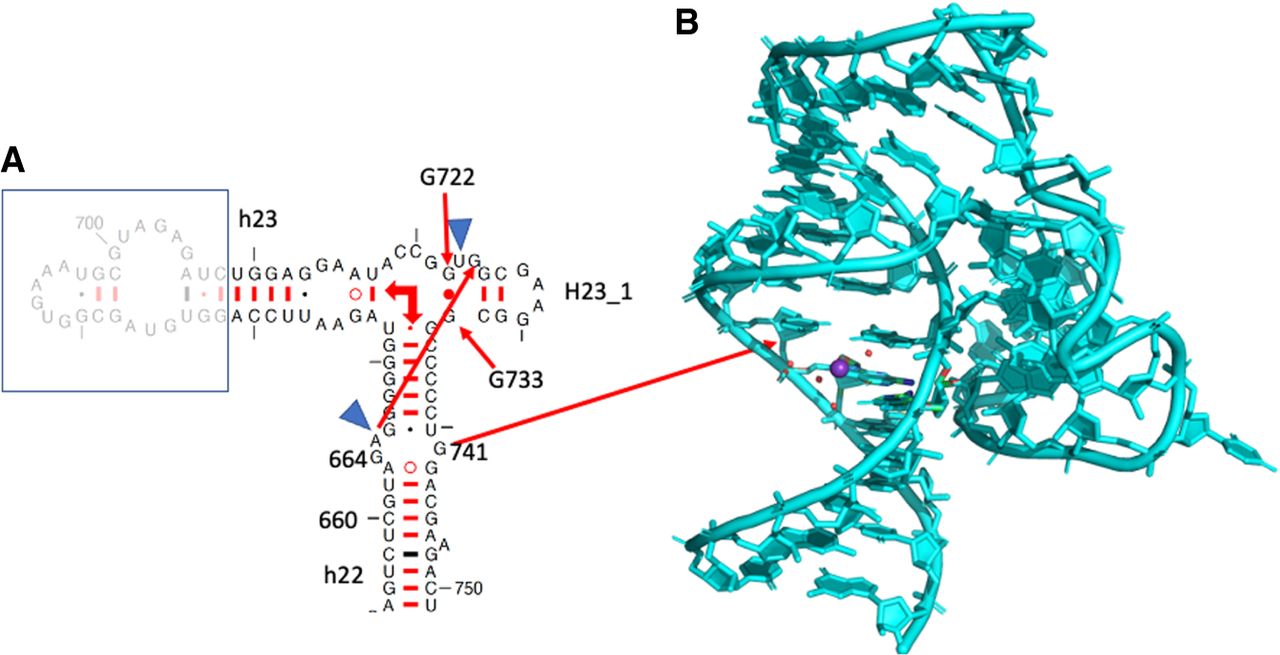
FIGURE 4.
(A) Part of the secondary structure of the 16S rRNA showing helices h22, h23, h23_1. The blue triangles indicate that those two residues bulge out so that A665 and G724 can form a cis Hoogsteen/Watson–Crick pair. The right-angle arrows indicate the coaxial stacking between the helices in the three-way junction. (B) The figure shows how the shallow minor groove side of the anionic G664•G741 binds tightly to the hairpin h23_1. Drawing based on E. coli (PDB 8B0X, Fromm et al. 2023).




