


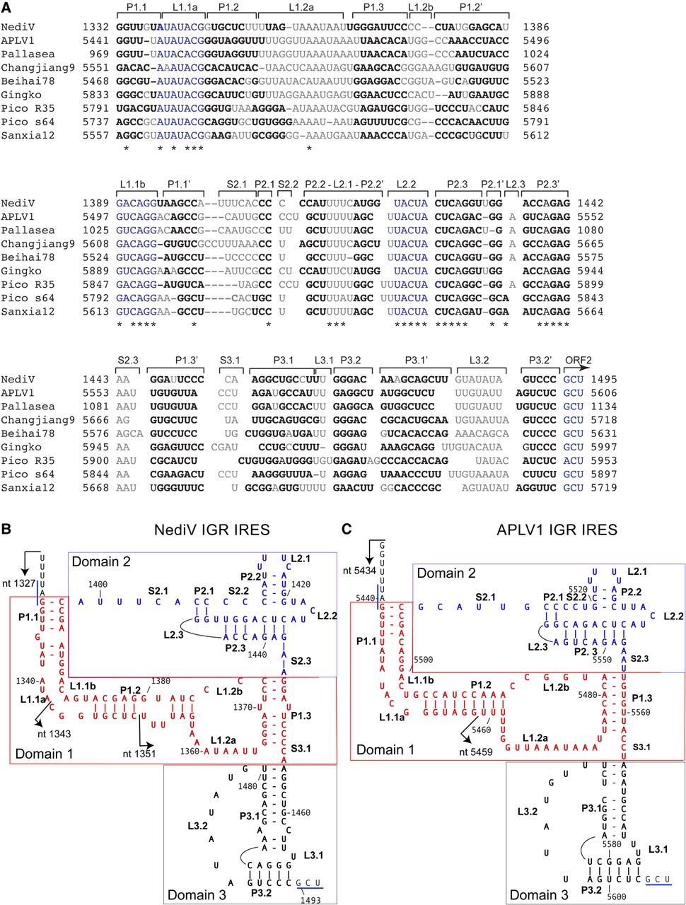
Sequence alignments and structural models of NediV and APLV1 IGR IRESs. (A) Aligned sequences from nedicistrovirus (NediV), Antarctic picorna-like virus 1 (APLV1), a viral TSA fragment from Pallasea cancelloides (Pallasea), Changjiang picorna-like virus 9 (Changjiang9), Beihai picorna-like virus 78 (Beihai78), Gingko biloba dicistrovirus (Gingko), Picornavirales sp. isolates R35-k141_316374 (Pico R35) and s64-k141_2464283 (Pico s64), and Sanxia picorna-like virus 12 (Sanxia12), annotated to show nucleotide numbers, structural elements (as in panels B,C), the ORF2 initiation codon, conserved sequence motifs (blue), base-paired nucleotides (bold), unpaired residues (light gray or light blue), and conserved nucleotides (indicated by asterisks). (B,C) Models of (B) NediV and (C) APLV1 IRESs. Nucleotides are numbered at 20-nt intervals. Domains 1 (PKII), 2 (PKIII), and 3 (PKI) are colored red, blue, and black, respectively. The 5′ borders of truncated IGR mRNAs used to define the IRES 5′ border (see Fig. 4A,B) are indicated by arrows. The NediV codon GCU1493–5 and the APLV1 codon GCU5604–6 that predominantly occupy the A site on initial binding of ribosomes, and the NediV UAG1330–2 and APLV1 UAG5439–41 ORF1 stop codons are indicated by blue lines.




