


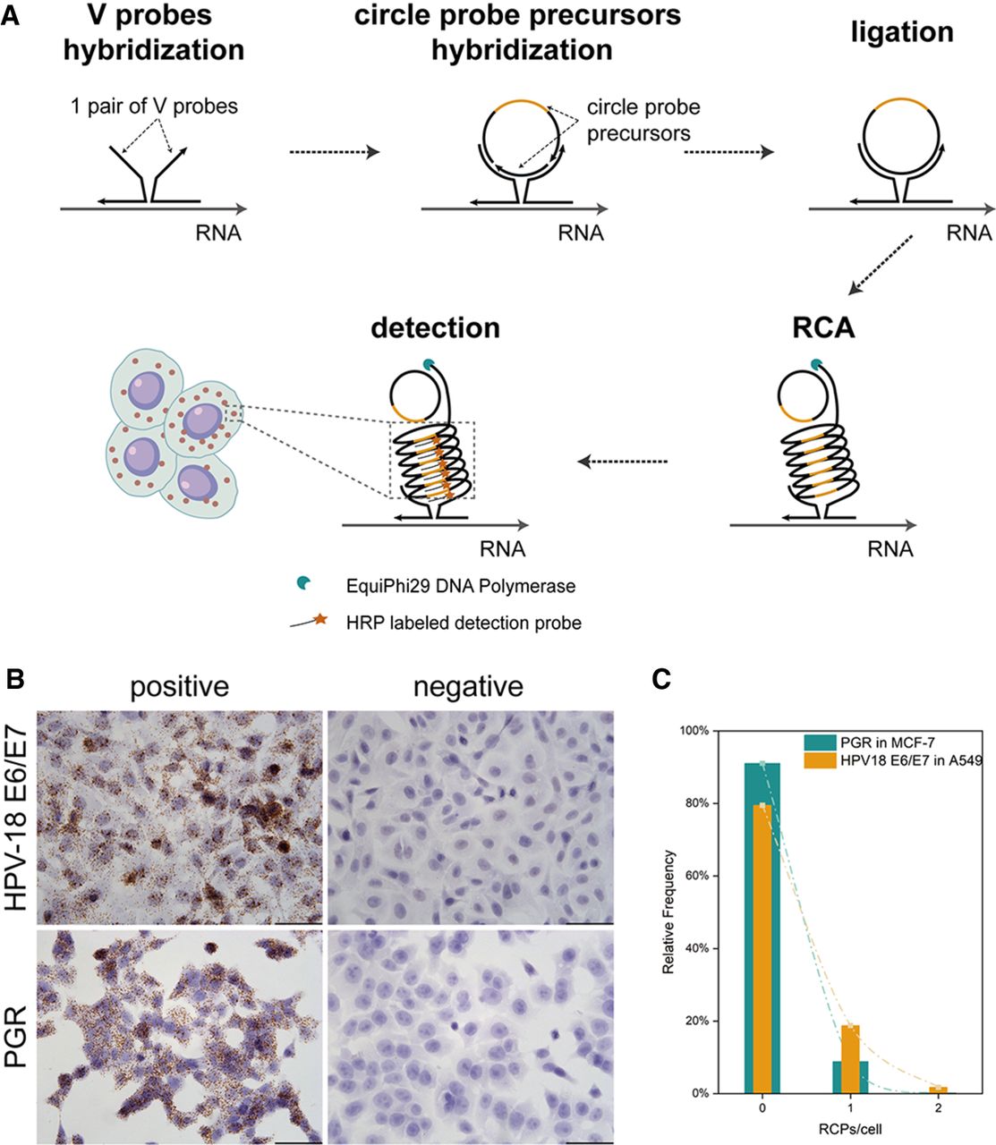
FIGURE 1.
Implementation of vsmCISH. (A) The workflow of vsmCISH. (B) Detection of HPV-18 E6/E7 expression in HeLa (positive) and A549 (negative) cells. Detection of PGR expression in T47D (positive) and MCF-7 (negative) cells. Scale bar, 50 µm. (C) Statistics plot of the relative frequency of cells without target gene expression.




