


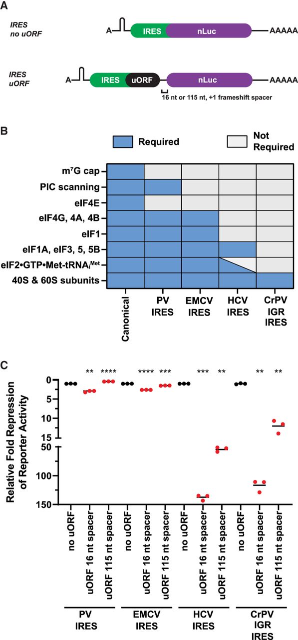
uORFs translated by IRESs that require less initiation factors permit less reinitiation. (A) Schematic of A-capped IRES-mediated reinitiation reporters. A stable hairpin was inserted upstream of the IRES to block scanning ribosomes. (B) Requirements of canonical initiation and class I–IV viral IRES-mediated initiation. (C) Response of canonical initiation and viral IRES-dependent nLuc reporters without and with small uORFs from in vitro translation. n = 3 biological replicates. Bar represents the mean. (PV) poliovirus, (EMCV) encephalomyocarditis virus, (HCV) hepatitis C virus, (CrPV IGR) cricket paralysis virus intergenic region, (PIC) preinitiation complex, (eIF) eukaryotic initiation factor. Luminescence signal of each IRES uORF reporter (in red) was set relative to their respective control IRES no uORF reporter (in black). Comparisons between each control IRES and their respective experimental IRES reporters were made using a two-tailed unpaired t-test with Welch's correction. (**) P < 0.01. (***) P < 0.001. (****) P < 0.0001. Exact P-values can be found in Supplemental Table 2.




