


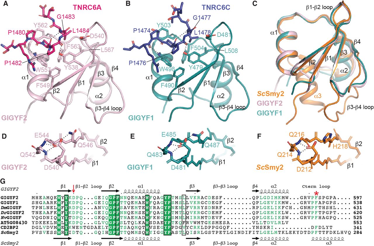
Overall structures of GIGYF1/2 GYF domains in complex with TNRC6 PPGL-containing peptides. (A) The TNRC6A PRS peptide (hot pink sticks) binds to a conserved arrangement of aromatic residues on the surface of the GIGYF2 GYF domain (light pink cartoon). Hydrogen bonds are denoted by dashed lines. Similar interactions are observed between the TNRC6C PRS peptide (navy sticks) and the GIGYF1 GYF domain (teal cartoon) (B). (C) The GIGYF1/2 GYF domains are highly similar to the ScSmy2 GYF fold (orange; PDB ID 3FMA [Ash et al. 2010]). (D–F) The conserved Asp residue that defines the Smy2 class of GYF domains mediates analogous interactions in GIGYF1/2 and ScSmy2. (G) Structural-based sequence alignment of GYF domains. The prototypical Asp residue of the Smy2 GYF subclass is indicated by the red arrow. The residues that comprise the PRS-binding surface are boxed in black. The Phe plug is denoted by the red asterisk.




