


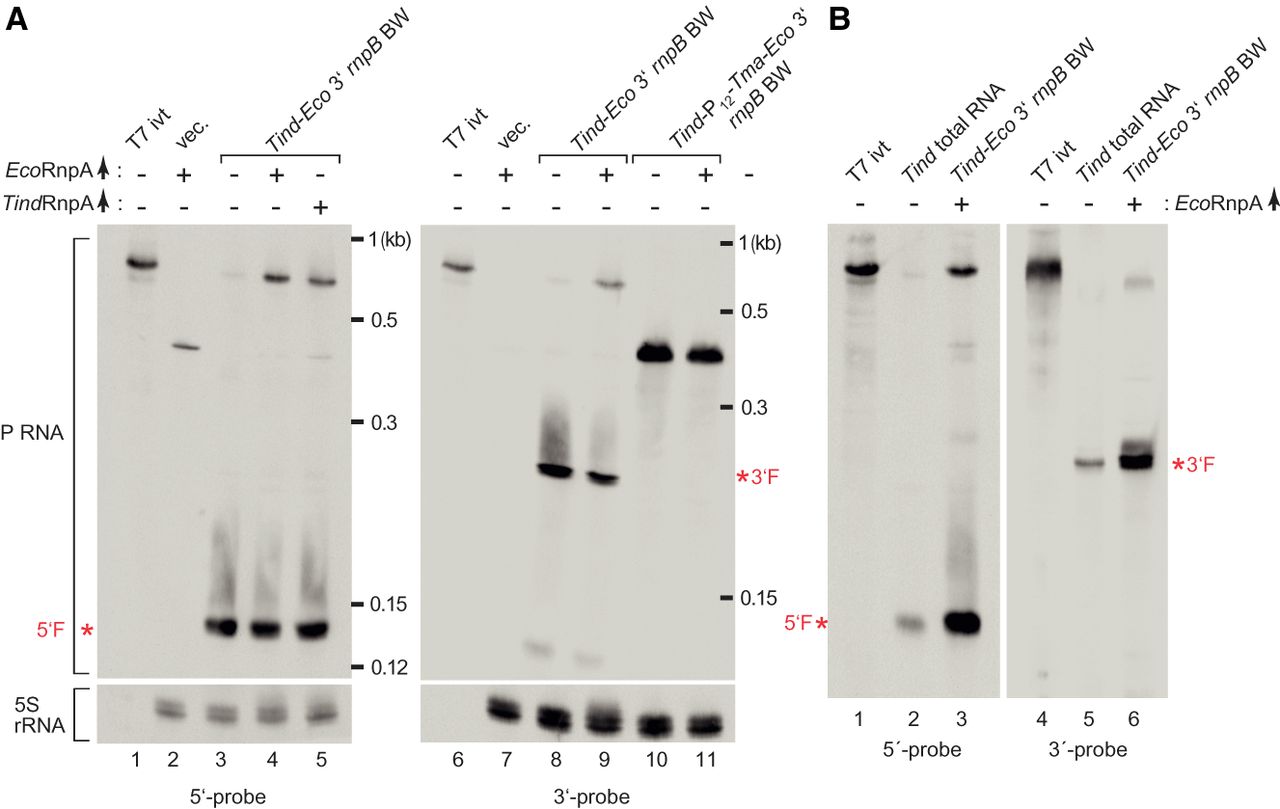
Northern blot analysis of T. indicus (Tind) P RNA expressed in E. coli BW or T. indicus. Total RNAs were isolated in exponential growth phase. DIG-labeled probes specific for the 5′- or 3′-region of Tind P RNA were complementary to nt 1–119 (5′-probe) and nt 312–339 (3′-probe) (see Fig. 1A; Supplemental Table S6). (A) Lanes “T7 ivt”: T7 in vitro transcript (see Supplemental Table S4) of Tind P RNA used as positive and length control; lanes “vec.”: total RNA extracted from E. coli BW transformed with the empty vector pACYC177. Simultaneous plasmid pBR322-borne overexpression of the E. coli or T. indicus RnpA protein in BW bacteria (indicated by EcoRnpA↑ or TindRnpA↑) is indicated above the lanes. The Tind P RNA variant Tind-P12-Tma- Eco 3′ (see Fig. 9) was expressed in lanes 10 and 11. The position and length (in kb) of bands of an RNA size marker (Low Range ssRNA Ladder, NEB) loaded onto the gels is indicated at the right margin of each blot. 5S rRNA was used as loading control. (B) Comparison of northern blot signals for Tind P RNA expressed in E. coli BW (lanes “Tind-Eco 3′ rnpB BW”) or in T. indicus cells (lanes “Tind total RNA”). For more details, see Materials and Methods. The northern blots shown in panels A and B are in each case representative examples out of three independent experiments. The signals corresponding to the in vivo-generated 5′-fragment(s) (5′F) and 3′-fragment(s) (3′F) of Tind P RNA are marked by asterisks and red lettering.




